Download
1 / 17
170 likes | 296 Views
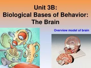
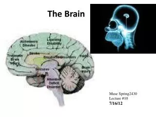
The Brain: Our Control Center. Chapter 3 Section 2. The Hindbrain. Medulla – vital function – heart rate, blood pressure, and breathing Pons – in the front of the Medulla regulate body movement, attention, sleep, and alertness. Cerebellum – (“little brain”) balance and coordination.

E N D
The Brain: Our Control Center Chapter 3 Section 2
The Hindbrain Medulla – vital function – heart rate, blood pressure, and breathing Pons – in the front of the Medulla regulate body movement, attention, sleep, and alertness. Cerebellum – (“little brain”) balance and coordination
The Midbrain Located between the hindbrain the forebrain area is involved in vision and hearing. Reticular Activating System- (begins in the hindbrain and rises to the forebrain) this system is important to attention, sleep, and arousal (increasing heart rate, blood pressure, and brain activity). Some drugs (alcohol) reduce the activity of the RAS. Loud noises can stimulate this system.
The forebrain makes it possible for humans to engage in complex thinking. Thalamus- (“inner chamber”) Relay station for sensory stimulation. Sense organs send information through the thalamus to the higher levels of the brain. Hypothalamus -(“under”) tiny area vital to regulating body temperature, storage of nutrients, areas of motivation and emotion, as well as, hunger, thirst, sexual behavior, caring for offspring and aggression. (disturbances lead to unusual eating or drinking behaviors. The Forebrain
Limbic system – forms a fringe along the inner edge of the cerebrum. It is involved in memory, emotion, hunger, sex, and aggression. Damage to specific parts can cause loss of new memories, aggressive or passive behaviors, etc. Cerebrum -(“Brain”)only human cerebrums make up such a large portion of the brain. 70% of the weight of the brain. Cerebral cortex- (“bark of a tree”)surface of the cerebrum that is wrinkled and has valleys. Deals with memory, language, emotions, complex motor functions, perception, etc. The Forebrain
Cerebral Cortex Two sides : left and right hemispheres (hemi- half)connected by the corpus callosum. Information received by one side of the body is transmitted to the opposite side’s hemisphere. Each side is divided into 4 lobes: Frontal lobe– behind the forehead Parietal lobe – to and rear of the head Temporal lobe – to the side just below the ears Occipital lobe- back of the head
Occipital Lobe – Primary visual area of the cerebral cortex. Damage to this lobe may cause people to recognize an object, but not be able to differentiate (facial recognition) Temporal Lobe- hearing or auditory area of the cortex Parietal lobe- messages received through the skin go through the sensory cortex in this lobe. Frontal lobe – motor cortex
Association areas- shape information into something meaningful. Left and Right Brains have many of the same functions, but differ in a number of ways. Most right handed people’s language function in the left brain (and many left handed) Wernicke’s Area (temporal lobe language function area)- pieces together sights and sounds damage makes it difficult to understand speech Boca’s Area (frontal lobe near the motor cortex)- control’s areas of the face used for speaking damage makes speaking difficult
Psychologists learn about left and right brain operations through split brain operations where the corpus callosum is cut, often in surgery’s for epilepsy. This procedure can reduce the severity of seizures, but the hemispheres cannot communicate with one another. (People can sometimes describe something held in their right hand, but not in their left)
Methods of Studying the Brain • Much of early research of the brain came from studying those with brain injury, now Psychologists use many methods
Accidents • http://www.youtube.com/watch?v=JTMg5YkxvYA • http://www.youtube.com/watch?v=TwwsrTznmVc • http://www.youtube.com/watch?v=sFME4dpBrOE&feature=related • http://www.youtube.com/watch?v=9Wl4-nNOGJ0 • Brain damage from head injuries can result in loss of vision, and hearing, confusion, or loss of memory. • Loss of large areas of brain may result in little loss of function, whereas loss of small vital areas a greater effect
Electrical Stimulation of the Brain • Electroencephalogram (EEG) • Scans • CATS • MRI • PET
EEG (Electroencephalogram)- a device that records the electrical activity of the brain. Used to diagnose some psychological disorders and to locate tumors through brain wave activity.
CAT (Computerized Axial Tomography)- x-ray beams are passed through the head by a moving ring. Density of brain tissue determines how much radiation is absorbed. The Computer them measures the amounts of radiation and pieces together a 3D view of the brain.
MRI (Magnetic Resonance Imaging ) – A person lies in a powerful magnetic field. Radio waves then cause parts of the brain to give off extra energy. The energy is measured at different angles and is translated into a visual image of the brain’s anatomy. MRI show details more clearly and is more powerful that a CT scan.
PET (Positron Emissional Tomography)- a person in injected with radioactive sugar. More of the radioactive sugar is used where brain activity is greater. A computer then uses sugar levels to create a image showing the amount of activity in different areas of the brain.