Download

1 / 16
160 likes | 342 Views
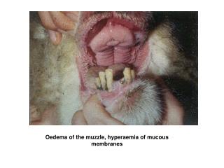
Mucous cysts of the DIPJ. Mucous cyst DIPJ. Ganglion cyst of the DIPJ Usually occurs between the fifth and seventh decades Associated with osteophytes or spurring of the DIPJ Osteoarthritis in other joints. Ganglion/Mucous cyst.

E N D
Mucous cyst DIPJ • Ganglion cyst of the DIPJ • Usually occurs between the fifth and seventh decades • Associated with osteophytes or spurring of the DIPJ • Osteoarthritis in other joints
Ganglion/Mucous cyst • Single or multiloculated cyst which appears smooth, white & translucent • Wall is made up of compressed collagen fibres and is sparsely lined with flattened cells without evidence of an epithelial or synovial lining • Mucin-filled “clefts” from the capsular attachment of the main cyst interconnect with the adjacent underlying joint via tortuous continuous ducts • Stroma may show tightly packed collagen fibres or sparsely cellular areas with broken fibres and mucin-filled intercellular & extracellular lakes • No inflammatory reaction or mitotic activity has been noted
Ganglion/Mucous cyst • Contents of cyst characterized by a highly viscous, clear, sticky, jelly-like mucin made up of glucosamine, albumin, globulin, & high concentrations of hyaluronic acid • Aetiology & pathogenesis remain obscure • Most widely accepted theory - mucoid degeneration associated with degeneration of joint capsule or tendon sheath • Injury & mechanical irritation may stimulate production of hyaluronic acid to form mucin, which may penetrate joint ligaments and capsules and then coalesce to form cyst
Clinical signs • Longitudinal grooving of the nail - earliest sign without a visible mass, caused by pressure on the nail matrix
Clinical signs • Enlarged cyst with attenuated overlying skin
Clinical signs • Cyst (3-5mm) usually lies to one side of the extensor tendon and between the dorsal distal joint crease & the eponychium
Clinical signs • Often has Heberden’s nodes and radiographic evidence of osteoarthritic changes in the joint
Treatment • Primarily surgical • Numerous alternative treatment reported in the past with moderate success: • Intralesional injection - eg. Sodium morrhuate, triamcinolone • Occlusive flurandrenolone tape
Surgical Management • Excision of the cyst alone • Wide excision of the cyst along with surrounding adjacent structures - eg.the overlying skin, osteophyte debridements • Debridement of the DIPJ osteophytes only, without excision of the cyst itself or overlying skin
Operative technique • L-shaped / H-shaped / curved incision • Elliptical excision of attenuated or involved skin
Operative technique • Cyst mobilized, traced to the joint capsule & excised with the joint capsule • All tissue excised between the extensor tendon & the adjacent collateral ligaments • Insertion of the extensor tendon and the nail matrix must be protected
Operative technique • Excison of osteophytes • Skin closure may require rotation / advancement dorsal skin flap or a full-thickness graft
Alternative approach • Transverse incision centred over DIPJ • Base of mucous cyst identified & excised while leaving the distal & superficial portion of the cyst intact • Excision of osteophtyes & joint capsule with direct skin closure • Allow several weeks for involution of the remaining cyst
Complications Residual nail deformities Stiffness Skin necrosis Recurrence: - inadequate excision - ganglion extension to the other side of extensor tendon - persistent underlying arthritic process