Download
1 / 26
260 likes | 486 Views
Joints. Rigid elements of the skeleton meet at joints or articulationsGreek root arthro" means jointArticulations can be:Bone to boneBone to cartilageTeeth in bony sockets. Classifications of Joints. Joints can be classified by function or structureFunctional classification based on amount

E N D
2. Joints Rigid elements of the skeleton meet at joints or articulations
Greek root �arthro� means joint
Articulations can be:
Bone to bone
Bone to cartilage
Teeth in bony sockets
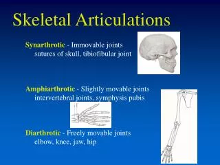
3. Classifications of Joints Joints can be classified by function or structure
Functional classification � based on amount of movement
Synarthroses � immovable � common in axial skeleton
Amphiarthroses � slightly movable � common in axial skeleton
Diarthroses � freely movable � common in appendicular skeleton
4. Classifications of Joints Structural classification based on:
Material that binds bones together
Presence or absence of a joint cavity
Structural classifications include:
Fibrous - CT fiber between bones
Cartilaginous - cartilage between bones
Synovial - joint cavity between bones
5. Fibrous Joints Bones are connected by fibrous connective tissue
Do not have a joint cavity
Most are immovable or only slightly movable
Types
Sutures - bones interlock (skull sutures)
Syndesmoses - bones do not interlock and are connected by ligaments (distal tibia / fibula)
6. Fibrous Joints
7. Cartilaginous Joints Bones are united by cartilage
Most are slightly movable (amphiarthrotic)
Lack a joint cavity
Two types
Synchondroses - bones united by hyaline cartilage (costal cartilages, epiphyseal plate)
Symphyses - bones connected by a flat disc of fibrocartilage (intervertebral discs and pubic symphasis)
8. Synchondroses
9. Symphyses Hyaline cartilage � also present as articular cartilage
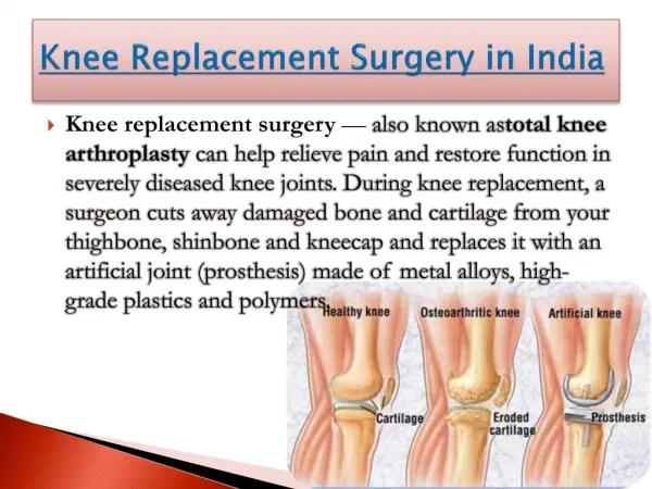
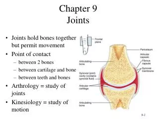
10. Synovial Joints Most movable type of joint
All are diarthroses
Each contains a fluid-filled joint cavity
Most are �simple� and have just two articulating surfaces
Some are �compound� with more than two articulating surfaces
Examples include the elbow (humerus, radius and ulna) and knee (femur, tibia, fibula, patella) joints
11. General Structure of Synovial Joints Articular cartilage
Ends of opposing bones are covered with hyaline cartilage
Absorbs compression
Joint cavity (synovial cavity)
Unique to synovial joints
Cavity is a potential space that holds a small amount of fluid (synovial fluid)
12. General Structure of Synovial Joints Articular capsule � joint cavity is enclosed in a two-layered capsule
Fibrous capsule � dense irregular connective tissue � strengthens joint
Synovial membrane � loose connective tissue
Lines joint capsule and covers internal joint surfaces
Functions to make synovial fluid
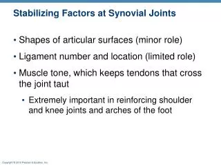
13. General Structure of Synovial Joints Reinforcing ligaments
Often are thickened parts of the fibrous capsule
Sometimes are extracapsular ligaments � located outside the capsule
Sometimes are intracapsular ligaments � located internal to the capsule
14. A Typical Synovial Joint
18. Sagittal Section of Knee Joint
19. Superior View of Knee Joint
20. Capsule of Knee Joint Covers posterior and lateral aspects of the knee
Covers tibial and femoral condyles
Does not cover the anterior aspect of the knee
Anteriorly � covered by three ligaments
Patellar ligament
Medial retinaculum
Lateral retinaculum
21. Anterior View of Knee
22. Knee Joint Extracapsular Ligaments
Become taut when knee is extended
Provide support of sides of the knee
Extracapsular ligaments:
Fibular (lateral) collateral ligament
Tibial (medial) collateral ligament
23. Anterior View of Knee
24. Knee Joint Intracapsular ligaments
Cruciate ligaments � cross each other like an �X�
Prevent undesirable movements at the knee joint
Each runs from the proximal tibia to the distal femur
Anterior cruciate ligament (anterior tibia to femur)
Posterior cruciate ligament (posterior tibia to femur)
25. Anterior View of Flexed Knee
26. Knee Joint Cruciate Ligaments
Prevent undesirable movements at the knee joint