Download
1 / 41
440 likes | 676 Views
The Heart. The Location of the Heart in the Thoracic Cavity. Figure 20.2c. The pericardia. Echocardiogram. The cardiovascular system is divided into two circuits. Pulmonary circuit blood to and from the lungs System circuit blood to and from the rest of the body

E N D
The Location of the Heart in the Thoracic Cavity Figure 20.2c
The cardiovascular system is divided into two circuits Pulmonary circuit blood to and from the lungs System circuit blood to and from the rest of the body Vessels carry the blood through the circuits Arteries carry blood away from the heart Veins carry blood to the heart Capillaries permit exchange
An Overview of the Cardiovascular System Figure 20.1
Compare (what is the same) and Contrast (what is different) Pulmonary and Systemic Circuits Oxygenated Deoxygenated Pressure Arteries Veins Color
Atria Thin walled chambers that receive blood from the vena cavae Ventricles Thick walled chambers separated from the atria by AV valves Chordaetendineae Tendinous fibers attached to the AV valves Papillary muscle and trabeculaecarneae Muscular projections on the inner wall of ventricles Internal Anatomy and Organization
Right atria Tricuspid valve Right ventricle Pulmonary valve Pulmonary circuit Left atria Bicuspid valve Left ventricle Aortic valve Aorta and systemic circuit Blood flow through the heart
The Sectional Anatomy of the Heart Animation: Diagrammatic Frontal Section through the Heart Figure 20.6a, b
The Sectional Anatomy of the Heart Figure 20.6c
Heart chambers and valves Structure and Function Question: Why does the human heart have four chambers?
Structural Differences in heart chambers The left side of the heart is more muscular than the right side Functions of valves AV valves prevent backflow of blood from the ventricles to the atria Semilunar valves prevent backflow into the ventricles from the pulmonary trunk and aorta Leaky valves???? Heart chambers and valves
Structural Differences between the Left and Right Ventricles • Volume • Thickness of wall • AV valve • Force of contraction Figure 20.7a-c
Valves of the Heart Figure 20.8a
Valves of the Heart Figure 20.8b
Components of the heart wall include Epicardium Myocardium Endocardium The Heart Wall
The Heart Wall and Cardiac Muscle Cells Figure 20.5a-c
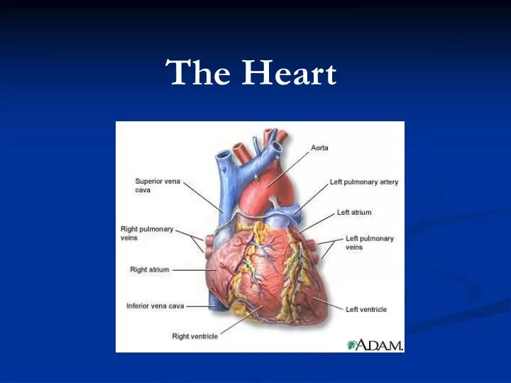
The heart consists of four chambers Two atria and two ventricles Major blood vessels of the heart include Inferior and superior vena cavae Aorta and pulmonary trunk Superficial Anatomy of the Heart
The Superficial Anatomy of the Heart Figure 20.3a
The Superficial Anatomy of the Heart Figure 20.3b, c
Arteries: right/ left coronary arteries, Veins: Great cardiac vein, anterior and posterior cardiac veins Blood Supply to the Heart
Coronary Circulation Figure 20.9c, d
The conducting system includes: Sinoatrial (SA) node Atrioventricular (AV) node Conducting cells Atrial conducting cells are found in internodal pathways Ventricular conducting cells consist of the AV bundle, bundle branches, and Purkinje fibers The Conducting System
SA node begins the action potential Stimulus spreads to the AV node Impulse is delayed at AV node Impulse then travels through ventricular conducting cells Then distributed by Purkinje fibers Impulse Conduction through the heart
Impulse Conduction through the Heart (figure 18.15) Figure 20.13
Cardiac Cycle Key Idea: Both atria fill an contract at same time Both ventricles fill and contract at the same time A Very efficient pump! Systole- contraction (ventricle) Diastole: relaxation (ventricle)
Regulation of Stroke Volume More stretch = more forceful contraction Think about rubber bands! High volume of venous return causes more stretching of myocytes Factors that increase venous return: 1. slow heart rate (why?) 2. exercise (why)
Regulation of Heart Rate Normal heart rate is _____________? Tachycardia and bradycardia…… Medulla Oblongata (Brain stem) Sympathetic Parasympathetic CardiaccelatoryCardioinhibitory Center (vagus nerve) center SA/AV node SA/AV node
Regulation of Heart Rate Sympathetic nervous system- speeds up heart rate epinephrine (adrenaline) , norepinephrine Stress and exercise, high level of calcium Parasympathetic nervous system- slows heart rate beta blockers, morphine- block epinephrine binding sites calcium channel blockers Alcohol- decreases activity of the vagus nerve- and the caridoaccelratory center in the brain stem.