Download
1 / 5
50 likes | 57 Views
Introduction: Spontaneous Osteonecrosis of the Knee (SONK) is a devastating and debilitating disease that mainly afflicts the elderly. A conservative approach may forgo the need for surgical intervention. This case report describes an orthopaedic patient diagnosed with SONK. After fi ve months of conservative treatment, the patient was able to walk without pain and remained clinically stable for seven years.

E N D
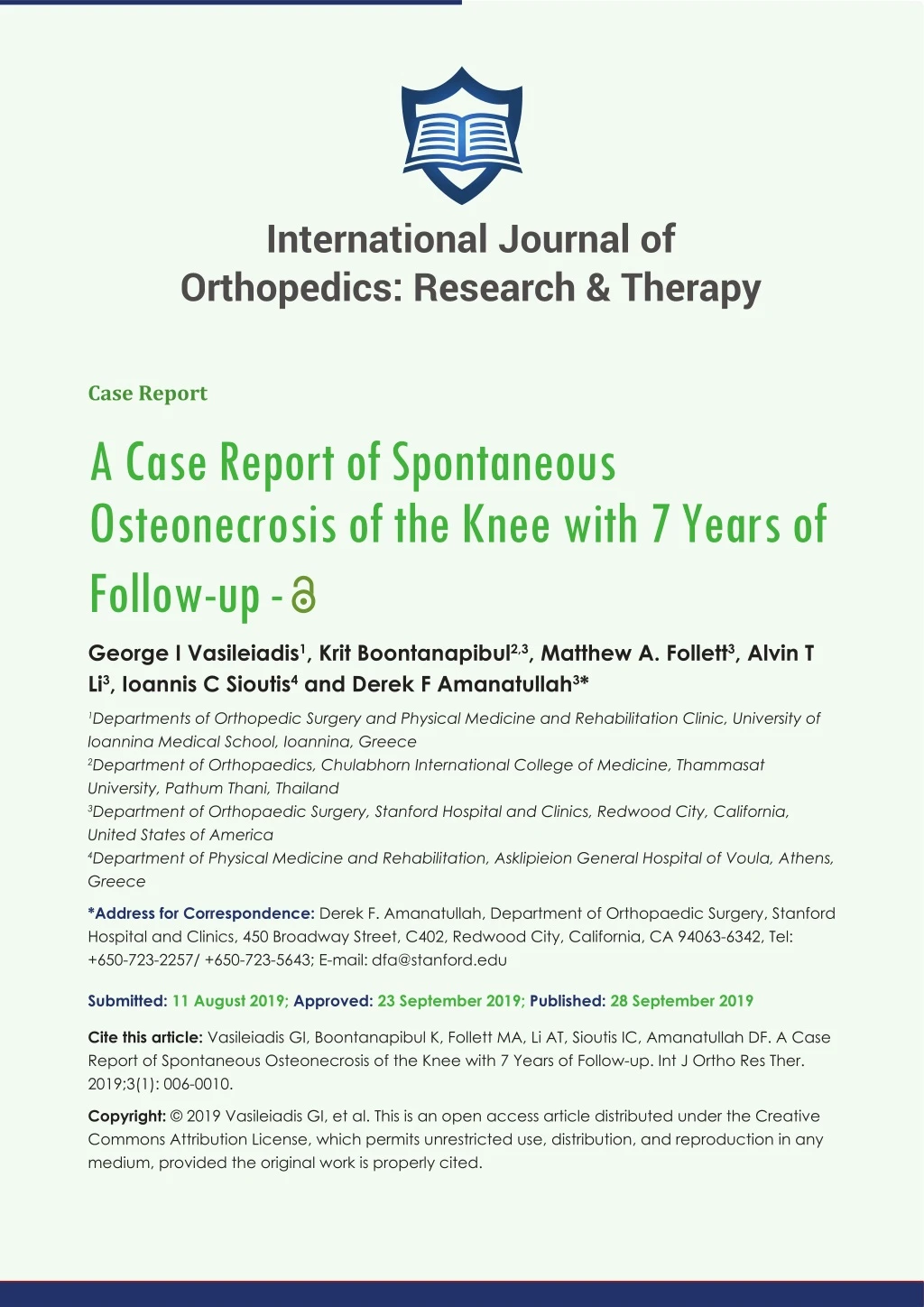
International Journal of Orthopedics: Research & Therapy Case Report A Case Report of Spontaneous Osteonecrosis of the Knee with 7 Years of Follow-up - George I Vasileiadis1, Krit Boontanapibul2,3, Matthew A. Follett3, Alvin T Li3, Ioannis C Sioutis4 and Derek F Amanatullah3* 1Departments of Orthopedic Surgery and Physical Medicine and Rehabilitation Clinic, University of Ioannina Medical School, Ioannina, Greece 2Department of Orthopaedics, Chulabhorn International College of Medicine, Thammasat University, Pathum Thani, Thailand 3Department of Orthopaedic Surgery, Stanford Hospital and Clinics, Redwood City, California, United States of America 4Department of Physical Medicine and Rehabilitation, Asklipieion General Hospital of Voula, Athens, Greece *Address for Correspondence: Derek F. Amanatullah, Department of Orthopaedic Surgery, Stanford Hospital and Clinics, 450 Broadway Street, C402, Redwood City, California, CA 94063-6342, Tel: +650-723-2257/ +650-723-5643; E-mail: Submitted: 11 August 2019; Approved: 23 September 2019; Published: 28 September 2019 Cite this article: Vasileiadis GI, Boontanapibul K, Follett MA, Li AT, Sioutis IC, Amanatullah DF. A Case Report of Spontaneous Osteonecrosis of the Knee with 7 Years of Follow-up. Int J Ortho Res Ther. 2019;3(1): 006-0010. Copyright: © 2019 Vasileiadis GI, et al. This is an open access article distributed under the Creative Commons Attribution License, which permits unrestricted use, distribution, and reproduction in any medium, provided the original work is properly cited.
International Journal of Orthopedics: Research & Therapy ABSTRACT Introduction: Spontaneous Osteonecrosis of the Knee (SONK) is a devastating and debilitating disease that mainly affl icts the elderly. A conservative approach may forgo the need for surgical intervention. This case report describes an orthopaedic patient diagnosed with SONK. After fi ve months of conservative treatment, the patient was able to walk without pain and remained clinically stable for seven years. Case Presentation: A 49-year-old male patient diagnosed with SONK of the left knee elected to undergo conservative management in lieu of surgical intervention. Seven years later the patient’s symptoms and MRI demonstrate a dramatic improvement. Conclusion: Current surgical procedures have had mixed results for treating SONK. All physicians, especially orthopedic surgeons, should be aware of expected outcomes for surgical intervention in SONK patients. Initial conservative management may be preferable to surgical intervention in some cases Keywords: Spontaneous osteonecrosis of the knee; SONK; Conservative treatment; Surgical treatment INTRODUCTION Osteonecrosis of the knee is a debilitating condition characterized by knee pain and bone marrow lesions, typically aff ecting both distal femoral condyles and/or the proximal tibia. It can be further delineated into two primary categories: Spontaneous Osteonecrosis of the Knee (SONK) and secondary osteonecrosis. SONK is particularly common in elderly females, aff ecting over 9% of patients over the age of 65 years [1-3]. SONK classically presents with acute onset of intense pain isolated to the medial femoral condyle [4]. Th e condition rarely occurs in lateral femoral condyle, tibial plateaus, and patella [5-7]. Additionally, eff usion and pain may limit range of motion [8,9]. Th e etiology of SONK is unclear, but it may originate from subchondral insuffi ciency or stress fracture [10,11]. In contrast, secondary osteonecrosis tends to present with gradually worsening pain in the medial and lateral condyles bilaterally, and other large joint involvement, particularly the hip [12]. Secondary osteonecrosis is typically found in patients under 55 years of age with risk factors including corticosteroid use, sickle-cell disease, alcohol consumption, Caisson’s disease, Gaucher’s disease, rheumatoid arthritis, and systemic lupus erythematosus [1,2]. In secondary osteonecrosis, ischemia and osteonecrosis lead to subchondral fracture, cartilage degeneration, and eventual subchondral collapse of the aff ected condyles [9,12]. condyle 3 to 4 days before he sought medical attention. Th is pain was intermittent during normal walking and aggravated by climbing up or down stairs. Th e patient reported no recent injury to his knee and is sedentary at home and at work. He has an unremarkable medical history, but smokes 20 cigarettes per day. On physical examination, the patient was afebrile with stable vital signs as well as a normal general appearance. Focused examination revealed slight eff usion of the left knee without noticeable muscular atrophy or skin discoloration. Palpation of the medial femoral condyle elicited pain radiating to the lateral femoral condyle, especially during knee fl exion. Th e knee was stable to varus and valgus stress at 30° of extension. Th e patient had a negative Lachman’s test, a negative posterior drawer, a negative dial test, as well as an intermittently positive McMurray’s test. Strength testing was normal at 5/5 for both knee fl exion and extension, but knee fl exion was restricted by pain. Sensation was intact to light touch in all nerve distributions. Th ere was a palpable dorsalis pedis pulse. No long track signs or distal edema were detected. Blood tests revealed no autoimmune or infectious disease. Radiographs of the knee were negative for gross involvement of the bone or misalignment (Figure 1). Aft er six weeks of analgesic and anti-infl ammatory therapy, symptoms persisted and worsened. At this time, an MRI was obtained (Figure 2). Available treatment options diff er between SONK and secondary osteonecrosis. In early stages or lesions smaller than 3.5 cm2, SONK has been successfully managed with conservative treatment [8,13]. Th is consists of protected weight bearing with crutches, analgesics, non-steroidal anti-infl ammatory drugs, and physical therapy. In refractory SONK and secondary osteonecrotic lesions larger than 5 cm2, necrotic segments may result in subchondral collapse or extension of the lesion [14,15]. Th is presentation of SONK, similar to secondary osteonecrosis, is oft en treated with invasive surgical procedures, including core decompression with or without bone graft ing, arthroscopy, osteochondral graft ing, high tibial osteotomy, or arthroplasty [2,16]. Th is case report describes an orthopaedic patient diagnosed with SONK. Aft er fi ve months of conservative treatment, the patient was able to walk without pain, and remains clinically stable for seven years. CASE PRESENTATION A 49-year-old man presented to the outpatient clinic on January 2009 complaining of excruciating pain of both the medial and lateral femoral condyles of his left knee. His pain began in the medial condyle one month prior, increasing in intensity and spreading to the lateral With a working diagnosis of SONK, conservative treatment was initiated with the patient’s consent. His treatment regimen consisted of crutch training to reducing weight bearing on the knee, physical therapy focused on strengthening the quadriceps and hamstring muscles, and non-steroidal anti-infl ammatory drugs. Aft er 5 months of conservative management, the patient’s symptoms had resolved completely. A follow-up MRI, 7 years later, revealed marked improvement in the osteonecrotic lesion (Figure 3). DISCUSSION Etiology and pathophysiology In spite of its prevalence, the etiology of SONK is poorly understood. No clear medical or genetic risk factors have been associated with SONK. SONK most frequently aff ects females over 65 years of age. Several possible causes have been proposed, including subchondral stress in osteopenic bone with insuffi ciency fractures [10,11,17] and meniscal tears [18-20]. Chronic or minor subchondral trauma in osteoporotic bone could induce microfractures, leading to fl uid accumulation in the intracondylar space, increasing intraosseous pressure, leading to vascular compromise, subsequent focal osseous SCIRES Literature - Volume 3 Issue 1 - www.scireslit.com Page -007
International Journal of Orthopedics: Research & Therapy DIAGNOSIS SONK can be diagnosed based on symptomatic and radiological criteria. Th e most characteristic symptoms include unilateral acute onset of pain isolated to the medial femoral condyle. Th e lateral condyle, tibial plateau, and patella are also susceptible. Small to moderate eff usion along with tenderness over the aff ected area is typical. Flexion contracture and decreased range of motion secondary to pain or eff usion may also be present. In contrast, secondary osteonecrosis tends to present with gradual onset of pain, and is frequently bilateral [9,22]. ischemia, and fi nally osteonecrosis [11]. Similarly, meniscal lesions have been associated with SONK, where loss of meniscal cartilage increases subchondral stress, leading to microfractures and necrotic bone in the same way [19,20]. SONK may not be a true osteonecrosis, as some authors have reported fi nding no necrotic tissue in aff ected condyles of patients diagnosed with SONK [3,20,21]. Plain radiograph is an inexpensive tool for initial evaluation and monitoring disease progression; however, it oft en does not show any abnormalities in the early stages of osteonecrosis [4,23]. Bone scintigraphy and Magnetic Resonance Imaging (MRI) are far more eff ective for diff erential diagnosis of osteonecrosis. Greyson et al. described a three-stage scintigraphic staging study for spontaneous osteonecrosis of the femoral condyle [24]. Following these criteria, markedly increased focal activity in all three phases is diagnostic for acute osteonecrosis. Normal activity in the fl ow-phase and increased activity in blood-pool and delayed phases is diagnostic for secondary osteonecrosis. MRI is the imaging modality of choice because of high sensitivity in detection of bone edema [25] and ability to evaluate meniscal and chondral pathology. SONK presents with an MRI pattern of subcortical focal loss of signal, along with homolateral meniscal degeneration or tearing, and focal deformity of the subchondral plate. No demarcation rim is typically observed. In contrast, secondary osteonecrosis frequently presents with the demarcation rim, and infrequent homolateral meniscal lesions and focal deformity of the subchondral bone plate [22]. Th erefore, MRI and bone scintigraphy allow diff erential diagnosis of osteonecrosis, distinguishing it from diff erent pathologies such as primary osteoarthritis, meniscal tears, and pes anserinus bursitis. Figure 1: Anterior to posterior radiograph of left knee at the time of presentation. A) B) In our case, the symptom that led the patient to seek medical attention was sudden excruciating pain in the left knee. Plain radiographs revealed no abnormalities. Th e fi rst abnormalities were found with an initial MRI, which revealed bone marrow edema and a minute fracture in the joint surface of the medial condyle. Subsequent MRI revealed enlargement of the fracture (2.2 x3 cm2). Figure 2: T1-weighted (A) and T2-weighted (B) MRI of left knee after six weeks of analgesic and non-steroidal anti-infl ammatory treatment. At this time, the patient reported that the pain had progressed and worsened. Treatment: Non-operative In asymptomatic patients, initial treatments for SONK and secondary osteonecrosis are similarly conservative [9]. Th erapy includes reduced weight-bearing using crutches or a walker, non- steroidal anti-infl ammatory drugs, and physical therapy focused on strengthening the quadriceps and hamstring muscles. Th e goal of conservative treatment is to prevent progression of the disease to subchondral collapse. Even in symptomatic patients, continuation of conservative treatment is eff ective for SONK patients. Prognosis of non-operative treatment depends on lesion size. Smaller lesions (less than 3.5 cm2) tend to resolve completely, while large lesions (greater than 5 cm2 or involving more than 40% of the condyle) tend to progress to subchondral collapse [14,15,26]. Treatment: Arthroscopic debridement Arthroscopy allows visualization of the aff ected areas, which is particularly useful for visualization when there is a question of lesion size or coexisting damage (e.g., meniscal tear). Miller et al. performed arthroscopic debridement on fi ve patients with idiopathic osteonecrosis. At a mean follow-up time of 31 months, four out of Figure 3: T1-weighted MRI of left knee at seven years follow-up after fi ve months of non-operative treatment. SCIRES Literature - Volume 3 Issue 1 - www.scireslit.com Page -008
International Journal of Orthopedics: Research & Therapy fi ve patients were rated good post-operatively based on the Hospital for Special Surgery Rating System. Th e average pre-operative score was 52 points and post-operative score was 82 points [27]. based on modifi ed Hospital for Special Surgery, modifi ed Cincinnati, Lysholm, and International Cartilage Repair society scores. Treatment: High tibial osteotomy Treatment: Core decompression High tibial osteotomy involves making a transverse cut in the proximal tibial metaphysis and removing a wedge of bone to change the geometry of the lower limb. Th is procedure seeks to change the geometry of the limb in order to reduce the load on the aff ected condyle. High tibial osteotomy has had promising results for SONK patients. Aglietti et al. followed 31 patients treated with high tibial osteotomy, 21 of which had also undergone ancillary bone graft ing. At a mean follow-up of 6.2 years, 87% of these patients had successful outcomes, and only two knees required arthroplasty [14]. Use of high tibial osteotomy is limited in secondary osteonecrosis because these patients tend to have tibial or bicondylar femoral involvement [3,9]. Th e principle behind core decompression is reduction of interosseous pressure via surgical extra-articular drilling of the aff ected condyle, promoting restoration of adequate circulation. Core decompression is a more conservative approach, which may delay the need for total knee arthroplasty. Forst et al. performed this procedure and reported immediate relief of pain in 15 out of 16 patients [28]. At 36 months follow-up, MRI confi rmed successful normalization of bone marrow signal. Th e authors recommended this procedure for early osteonecrosis prior to fl attening of the femoral condyle. In secondary osteonecrosis, core decompression is similarly eff ective in early stages of the disease. Mont et al. compared core decompression with non-operative treatment in 79 knees. Th ey performed core decompression on 47 knees with Ficat and Arlet stage I to stage III secondary osteonecrosis. At a mean follow-up time of 11 years, 73% of patients had good to excellent outcomes based on Knee Society Scores of 80 points or greater. Radiographs showed progression to Ficat and Arlet stage III or IV in 36% of core decompression knees, as opposed to 75% of nonoperative knees [29]. Treatment: Unicondylar arthroplasty Unicondylar arthroplasty has been used with success in SONK due to the tendency of the disease to be confi ned to one condyle. Th is procedure involves replacing the aff ected femoral condyle and associated tibial articular surface. In contrast, this procedure is not generally recommended for secondary osteonecrosis if it involves both condyles. In a study of 41 SONK patients, Chalmers et al. reported an 93% success rate in unicondylar knee arthroplasty for an isolated compartment at both fi ve and ten years follow-up [33]. Th is treatment has the advantage of rapid post-operational recovery and minimizing eff ects on the cruciate ligaments, patella, and the other compartment of the knee. A recent meta-analysis study also reported that cemented medial unicondylar knee arthroplasty has the same clinical and survival outcome as medial compartment knee osteoarthritis when treating SONK [34]. In a similar procedure, Goodman and Hwang investigated the effi cacy of local debridement with osteoprogenitor cell graft ing in twelve patients with secondary osteonecrosis of the distal femoral condyle. Th e premise of the study was to correct the biological defi ciency of live osteoprogenitor cells in the subchondral and metaphyseal areas of necrotic femoral condyle. Th is procedure involved open debridement of the osteonecrotic lesion and drilling in at multiple angles, excising easily accessible loose necrotic bone. Th e defect was then fi lled with concentrated bone marrow osteoprogenitor cells harvested from the iliac crest. At an average follow-up time of 5 years, Knee Society Score averaged 87 points, and Knee Function Score averaged 85 points. Th e investigators recommended this procedure for young patients with secondary osteonecrosis of the knee prior to collapse of the femoral condyle [30]. Treatment: Total knee arthroplasty Total knee arthroplasty is reserved for late-stage osteonecrosis when patients are in severe pain that has not responded to other interventions. It is indicated for late-stage secondary osteonecrosis with degenerative changes, patients with severe pain, and those with functional disability. Th is procedure involves replacing the patient’s femoral and tibial articular surfaces. Myers et al. reviewed the outcomes of total knee arthroplasty on 148 knees with SONK and 150 knees with secondary osteonecrosis. Th ey reported successful outcomes in 92% of SONK cases and 74% of secondary osteonecrosis cases based on Knee Society and Hospital for Special Surgery scores [16]. CONCLUSION We describe a case of successful conservative treatment of SONK. In spite of improvements in technique since 1985, it is clear that surgical treatment of osteonecrosis is not always met with success. It is important to diff erentiate between SONK and secondary osteonecrosis. Regarding the medical therapy of osteonecrosis of the knee, treatment is similar for both SONK and secondary osteonecrosis as long as the patient is asymptomatic. Th is encompasses a conservative regimen of partial weight bearing with crutches or a walker, non-steroidal anti-infl ammatory medications, and physical therapy focusing on strengthening the quadriceps and hamstrings. However, when the patient is symptoms, treatment options diff er. Conservative measures should be the fi rst consideration for SONK to minimizing invasive procedures and optimize long-term outcomes for patients. Treatment: Osteochondral allografts and autologous osteochondral transplantation Osteochondral repair may be used for patients for whom conservative treatment has failed. In the past, Bayne et al. attempted allograft s for 6 SONK patients and 3 corticosteroid- induced secondary osteonecrosis patients. Of these, no secondary osteonecrosis graft succeeded and only one SONK graft was successful [31]. Th e authors suspected that continued use of corticosteroids led to poor vascularization of the graft and subsequent subsidence in the secondary graft procedures. For SONK, it was hypothesized that the poor compliance of elderly patients resulted in allograft fragmentation. A more promising alternative has been autologous osteochondral transplantation. Th is treatment relies on transplanting articular cartilage with subchondral bone from less weight-bearing areas to the aff ected areas. Th e goal of osteochondral transplantation is to fi ll the aff ected area with cylindrical osteochondral allograft s to form a congruent hyaline cartilage covered surface. Hangody et al. reported good to excellent outcomes in 92% of 789 patients treated with femoral condylar implants [32]. Th eir results were calculated SCIRES Literature - Volume 3 Issue 1 - www.scireslit.com Page -009
International Journal of Orthopedics: Research & Therapy CLINICAL MESSAGE Th e current surgical procedures available for treating SONK have mixed results. All physicians, especially orthopedic surgeons, should be aware of the expected outcomes for surgical intervention in patients with SONK. Initial conservative management may be preferable to surgical intervention in some cases. ACKNOWLEDGEMENTS AND FUNDING No outside funding was required to conduct this case report. Additionally, this case report was Institutional Review Board (IRB) exempt. REFERENCES osteonecrosis of the knee. J Bone Joint Surg Am. 2006; 88: 76-82. http://bit. ly/2mfTfi o 17. Akamatsu Y, Mitsugi N, Hayashi T, Kobayashi H, Saito T. Low bone mineral density is associated with the onset of spontaneous osteonecrosis of the knee. Acta Orthop. 2012; 83: 249-255. http://bit.ly/2ldytji 18. Brahme SK, Fox JM, Ferkel RD, Friedman MJ, Flannigan BD, Resnick DL. Osteonecrosis of the knee after arthroscopic surgery: diagnosis with MR imaging. Radiology. 1991; 178: 851-853. http://bit.ly/2mJTZg2 19. Robertson DD, Armfi eld DR, Towers JD, Irrgang JJ, Maloney WJ, Harner CD. Meniscal root injury and spontaneous osteonecrosis of the knee: an observation. J Bone Joint Surg Br. 2009; 91: 190-195. http://bit.ly/2mLgJw9 20. Yasuda T, Ota S, Fujita S, Onishi E, Iwaki K, Yamamoto H. Association between medial meniscus extrusion and spontaneous osteonecrosis of the knee. Int J Rheum Dis. 2018; 21: 2104-2111. http://bit.ly/2mf5iN2 1. Pape D, Seil R, Fritsch E, Rupp S, Kohn D. Prevalence of spontaneous osteonecrosis of the medial femoral condyle in elderly patients. Knee Surg Sports Traumatol Arthrosc. 2002; 10: 233-240. http://bit.ly/2mffx3W 21. Takeda M, Higuchi H, Kimura M, Kobayashi Y, Terauchi M, Takagishi K. Spontaneous osteonecrosis of the knee: histopathological differences between early and progressive cases. J Bone Joint Surg Br. 2008; 90: 324- 329. http://bit.ly/2kMUc1q 2. Karim AR, Cherian JJ, Jauregui JJ, Pierce T, Mont MA. Osteonecrosis of the knee: review. Ann Transl Med. 2015; 3: 6. http://bit.ly/2kHEgxd 22. Narvaez J, Narvaez JA, Rodriguez-Moreno J, Roig-Escofet D. Osteonecrosis of the knee: differences among idiopathic and secondary types. Rheumatology (Oxford). 2000; 39: 982-989. http://bit.ly/2l0X9LX 3. Kattapuram TM, Kattapuram SV. Spontaneous osteonecrosis of the knee. Eur J Radiol. 2008; 67: 42-48. http://bit.ly/2lgT1aH 4. al-Rowaih A, Bjorkengren A, Egund N, Lindstrand A, Wingstrand H, Thorngren KG. Size of osteonecrosis of the knee. Clin Orthop Relat Res. 1993; 287: 68- 75. http://bit.ly/2kHBzvB 23. Pollack MS, Dalinka MK, Kressel HY, Lotke PA, Spritzer CE. Magnetic resonance imaging in the evaluation of suspected osteonecrosis of the knee. Skeletal Radiol. 1987; 16: 121-127. http://bit.ly/2lfqamX 5. Lotke PA, Abend JA, Ecker ML. The treatment of osteonecrosis of the medial femoral condyle. Clin Orthop Relat Res. 1982; 171: 109-116. http://bit. ly/2lfVMJb 24. Greyson ND, Lotem MM, Gross AE, Houpt JB. Radionuclide evaluation of spontaneous femoral osteonecrosis. Radiology. 1982; 142: 729-735. http:// bit.ly/2mKNDNq 6. LaPrade RF, Noffsinger MA. Idiopathic osteonecrosis of the patella: an unusual cause of pain in the knee. A case report. J Bone Joint Surg Am. 1990; 72: 1414-1418. http://bit.ly/2mfMfSG 25. Fotiadou A, Karantanas A. Acute nontraumatic adult knee pain: the role of MR imaging. Radiol Med. 2009; 114: 437-447. http://bit.ly/2l4poJJ 26. Juréus J, Lindstrand A, Geijer M, Robertsson O, Tägil M. The natural course of Spontaneous Osteonecrosis of the Knee (SPONK): a 1- to 27-year follow- up of 40 patients. Acta Orthop. 2013; 84: 410-414. http://bit.ly/2mLLD7L 7. Ohdera T, Miyagi S, Tokunaga M, Yoshimoto E, Matsuda S, Ikari H. Spontaneous osteonecrosis of the lateral femoral condyle of the knee: a report of 11 cases. Arch Orthop Trauma Surg. 2008; 128: 825-831. http://bit. ly/2mDHbHR 27. Miller GK, Maylahn DJ, Drennan DB. The treatment of idiopathic osteonecrosis of the medial femoral condyle with arthroscopic debridement. Arthroscopy. 1986; 2: 21-29. http://bit.ly/2kHRH09 8. Yates PJ, Calder JD, Stranks GJ, Conn KS, Peppercorn D, Thomas NP. Early MRI diagnosis and non-surgical management of spontaneous osteonecrosis of the knee. Knee. 2007; 14: 112-116. http://bit.ly/2lgOzsv 28. Forst J, Forst R, Heller KD, Adam G. Spontaneous osteonecrosis of the femoral condyle: causal treatment by early core decompression. Arch Orthop Trauma Surg. 1998; 117: 18-22. http://bit.ly/2mkLk3c 9. Zywiel MG, McGrath MS, Seyler TM, Marker DR, Bonutti PM, Mont MA. Osteonecrosis of the knee: a review of three disorders. Orthop Clin North Am. 2009; 40: 193-211. http://bit.ly/2kHLEZw 29. Mont MA, Tomek IM, Hungerford DS. Core decompression for avascular necrosis of the distal femur: long term followup. Clin Orthop Relat Res. 1997; 334: 124-130. http://bit.ly/2mhvSF9 10. Mears SC, McCarthy EF, Jones LC, Hungerford DS, Mont MA. Characterization and pathological characteristics of spontaneous osteonecrosis of the knee. The Iowa orthopaedic journal. 2009; 29: 38-42. http://bit.ly/2l3zevo 30. Goodman SB, Hwang KL. Treatment of secondary osteonecrosis of the knee with local debridement and osteoprogenitor cell grafting. J Arthroplasty. 2015; 30: 1892-1896. http://bit.ly/2mhvt5B 11. Yamamoto T, Bullough PG. Spontaneous osteonecrosis of the knee: the result of subchondral insuffi ciency fracture. J Bone Joint Surg Am. 2000; 82: 858-866. http://bit.ly/2mmIGtN 31. Bayne O, Langer F, Pritzker KP, Houpt J, Gross AE. Osteochondral allografts in the treatment of osteonecrosis of the knee. Orthop Clin North Am. 1985; 16: 727-740. http://bit.ly/2lcH8Cz 12. Mont MA, Baumgarten KM, Rifai A, Bluemke DA, Jones LC, Hungerford DS. A traumatic osteonecrosis of the knee. J Bone Joint Surg Am. 2000; 82: 1279- 1290. http://bit.ly/2mkUvAG 32. Hangody L, Vasarhelyi G, Hangody LR, Sukosd Z, Tibay G, Bartha L, et al. Autologous osteochondral grafting--technique and long-term results. Injury. 2008; 39: S32-39. http://bit.ly/2mKKAEY 13. Woehnl ANQ, Costa C. Osteonecrosis of the knee. Orthopaedic Knowledge Online Journal. 2012: 10. 33. Chalmers BP, Mehrotra KG, Sierra RJ, Pagnano MW, Taunton MJ, Abdel MP. Reliable outcomes and survivorship of unicompartmental knee arthroplasty for isolated compartment osteonecrosis. Bone Joint J. 2018; 100: 450-454. http://bit.ly/2mFvEI1 14. Aglietti P, Insall JN, Buzzi R, Deschamps G. Idiopathic osteonecrosis of the knee. Aetiology, prognosis and treatment. J Bone Joint Surg Br. 1983; 65: 588-597. http://bit.ly/2mm1mtC 15. Mont MA, Marker DR, Zywiel MG, Carrino JA. Osteonecrosis of the knee and related conditions. J Am Acad Orthop Surg. 2011; 19: 482-494. http://bit. ly/2mkhD2A 34. Yoon C, Chang MJ, Chang CB, Choi JH, Lee SA, Kang SB. Does unicompartmental knee arthroplasty have worse outcomes in spontaneous osteonecrosis of the knee than in medial compartment osteoarthritis? A systematic review and meta-analysis. Arch Orthop Trauma Surg. 2019; 139: 393-403. http://bit.ly/2mKIPrm 16. Myers TG, Cui Q, Kuskowski M, Mihalko WM, Saleh KJ. Outcomes of total and unicompartmental knee arthroplasty for secondary and spontaneous SCIRES Literature - Volume 3 Issue 1 - www.scireslit.com Page -0010