Download
1 / 66
840 likes | 1.4k Views
MEDIASTINAL DISEASES. By Abdellah Hamed MD, PhD (Japan) Ass. Prof of Respirology Sohag University. Anatomical Consideration. Anatomy of the Mediastinum. Mediastinum is the central space within the thoracic cavity bounded by: Sternum anteriorly Lungs and parietal pleura laterally

E N D
MEDIASTINAL DISEASES By Abdellah Hamed MD, PhD (Japan) Ass. Prof of Respirology Sohag University
Anatomy of the Mediastinum • Mediastinum is the central space within the thoracic cavity bounded by: • Sternum anteriorly • Lungs and parietal pleura laterally • The vertebral column posteriorly • The thoracic inlet superiorly • The diaphragm inferiorly
Divisions of the mediastinum : • Superior mediastinum: it contains • Aortic arch & its 3 branches • S.V.C. & its 2 innominate veins • Trachea, esophagus, thoracic duct • Vagus, phrenic n., left recurrent laryngeal n. • and sympathetic n. • L.N. & thymus.
Cont…. • (2)Anterior mediastinum : • Boundary: Anterior Sternum • Posterior Pericardium • Contents: Thymus • L.N. • Fatty tissue
Cont…. • (3)Middle mediastinum: • Boundary : By the 3 divisions. • Contents : Heart & pericardium • Ascending aorta, S.V.C & I.V.C. • Pulmonary arteries & veins • Tracheal bifurcation • Phrenic nerves
Cont…. • (4)Posterior mediastinum : • Boundary : Anterior pericardium & diaphragm • Posterior lower 8 thoracic vertebrae • Contents : Descending aorta • Esophagus • Sympathetic & vagus nerves • Thoracic duct • L.N.
Mediastinal Pathologies • Non neoplastic diseases • Mediastinitis • Pneumomediastinum • Congenital pathologies • Cysts • Hernias • Acquired lesions • Benign • Malignant
Types and sites of mediastinal lesions • Superior mediastinum : • Thymic tumors • Intrathoracic thyroid • Teratoma • Esophageal lesions • Cystic hygroma • Lymphomata • Mediastinal abscess
Cont…. • (B)Anterior mediastinum : • Thymic tumors & cysts • Teratoma • Intrathoracic thyroid • Cystic hygroma • Pleuro-pericardial cyst • Lymphomata
Cont…. • (C)Middle mediastinum : • Aortic aneurysm • Anomalies of great vessels • Bronchogenic cyst • Lipoma
Cont…. • (d)Posterior mediastinum: • Neurogenic tumors • Gastroenteric & bronchogenic cysts • Esophageal lesions • Meningocele • Aortic aneurysm • Cold abscess • Hernia through foramina of Bochdalek
Mediastinal Syndrome • This is usually results from compression of the mediastinal • structures by a mediastinal lesion. • Causes : • Mediastinal tumors • Chronic mediastinitis • Mediastinal emphysema
Malignancy Lung cancer Lymphoma Thymoma Metastatic Germ Cell “Benign” Infection/Inflammation Benign Neoplasms Iatrogenic Trauma Etiology of SVC
Malignancy • Account for 80-97% of SVCS cases • Lung Cancer 75-80% • Lymphoma 10-15% • Others 5% • Metastatic • Thymoma • Germ cell tumor Markman, M. Cleveland Clin JOM, 1999. Ostler, P. Clin Onc, 1997.
Cont…. Primary location of specific neoplasma & cysts within the subdivisions of the mediastinum
Cont…. Manifestations : 1) Pressure symptoms 2) Hormonal effects These depend on : * Site of lesion * Structure involved
Cont…. • Pressure symptoms : • Esophagus: dysphagia. • Trachea & bronchi : brassy cough, stridor, obstructive • emphysema or atelectasis • Arteries : unequal pulse, ischaemic manifestations • ( pallor, pain and syncope ).
Cont…. • Veins : usually S.V.Cdistension of neck veins, collaterals. • Nerves : * Sympathetic Horner’s syndrome . • * Vagus dysphagia & arrhythmia . • * Recurrent laryngeal hoarseness of voice . • * Phrenic diaphragmatic paralysis.
Cont…. • Hormonal : • Retrosternal goiterToxic changes • Thymic tumor Myasthenia gravis • Parathyroid adenoma Hyperparathyroidism
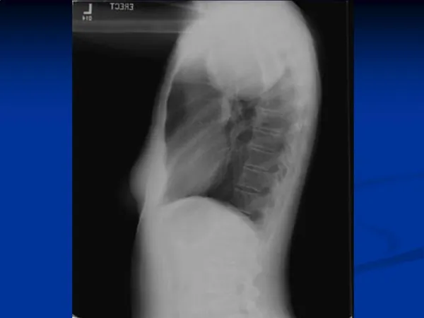
Diagnostic Procedures • Chest radiograph • Duplex ultrasound • CT/MRI/MRV • Venogram • Radionuclide studies • Thoracoscopy or thoracotomy
Computed tomography Can identify normal anatomic variations and fluid filled cyst Site of the origin of the mass can be better identified 100% specificity for the CT appearance of teratomas, thymolipoma, omental fat herniation Overall accuracy for predicting mediastinal mass is only 48%
Computed tomography • Limitation • Horizontal oriented structures and boundaries are difficult to evaluate • Abnormalities in the aortopulmonary window area and the subcarinal area • CT has become the initial imaging procedure of choice for evaluation of mediastinum in patients with primary mediastinal mass or with lung cancer
Magnetic Resonance Imaging Assesses tissue by measuring the radiofrequency induced nuclear resonance instead of measuring the attenuation of transmitted ionizing radiation Coronal and sagittal planes are better viewed, vertical structures and boundaries are better evaluated Superior sulcus tumors, lesions invading the medistinum, chest wall and diaphragm And possible invasion of the brachial plexus, and for evaluating vertebral invasion
Magnetic Resonance Imaging • Limitations • Distinguish poorly between hilar mass and adjacent collapsed or consolidated lung • Cannot distinguish between a benign and a malignant causes for lymph node enlargement
Ultrasonography For cystic nature of mediatinal mass Useful in guiding endoscopic biopsy technique
Radionuclide imaging Rely on the localization of markers based on specific metabolic or immunologic properties of the target tissue Potential ability to diagnose and stage a malignancy and identify distant metastasis Planar imaging with gallium 67 and thallium-201
POSITRON EMISSION TOMOGRAPHY The technique is not infallible because certain non-neoplastic processes, including granulomatous and other inflammatory diseases as well as infections, may also demonstrate positive PET imaging Size limitations are also an issue, with the lower limit of resolution of the study being approximately 7 to 8 mm depending on the intensity of uptake of the isotope in abnormal cells One should not rely on a negative PET finding for lesions less than 1 cm on CT scan
ENDOSCOPIC ULTRASOUND Superior ability to sample the posterior mediastinum through the esophageal wall For patients with lung cancer and posterior mediastinal adenopathy seen on chest CT scan EUS has a sensitivity and specificity of 90% and 100%, respectively.
Mediastinoscopy Allows direct inspection and biopsy of lymph nodes or other masses on the superior portion of the anterior mediastinum
MEDIASTINOSCOPY • Mediastinoscopy remains the gold standard for invasively staging the mediastinum • If there is mediastinal adenopathy on CT, often a surgical mediastinal procedure is performed • Mediastinoscopy is most often used to sample lymph nodes in the • Paratracheal (station 4) • Anterior subcarinal (station 7) • The subcarinal area is more difficult to sample and thus has a lower yield
Venography • Can give precise level of obstruction • Less information on etiology of SVCS • Requires larger contrast dose
Tissue Diagnosis ProcedureYield Sputum cytology 33-40% Bronchoscopy 33-60% LN biopsy 46-80% Mediastinoscopy 100% Thoracotomy 100% Ostler, J. Clin Onc, 1997 Schindler, N. Surg Clin N Am, 1999
Which First---> Tx or Dx? • Ahman • Literature search 1934-1984 • 1986 cases SVC reviewed • Only 1 clearly documented death 2/2 SVCS Ahman, F. J Clin Onc, 1984.
Treatment • Tailored to etiology • Historically standard tx----->XRT • Emergent tx before tissue dx 2/2 presumed risk of bleeding • Current standard----> tissue dx prior to initiating tx
Treatment • Goal • treat symptoms • treat underlying cause • Tx should be tailored to histologic diagnosis---->determine if curative vs palliative
Treatment • Chemotherapy • XRT • Surgery • Interventional Procedures Spiro, S. Thorax, 1983 Perez-Soler, P. J Clin Onc, 1984
Prognosis • Varies depending on the etiology • SVCS in its own right is rarely fatal • 10-20% survive at least 2 years Ahman,F. J Clin Onc, 1984 Ostler, PJ. Clin Onc, 1997 Perez & Brady, 2004.
Non neoplastic Disorders of the Mediastinum • Pneumomediastinum • Pneumopericardium • Acute Mediastinitis • Chronic Mediastinitis
Acute Mediastinitis • Causes: • Esophageal perforation: • * Traumatic : endoscopies, dilatation, intubations • * Spontaneous • Operation : in the larynx, trachea, esophagus • Suppurative L.N. secondary to infection of the lung, • esophagus & larynx.
Cont…. • T.B, osteomyelitis of cervical or thoracic spine. • Direct extension of infection from the • neck, retropharyneal space, pleura, pericardium.
Cont…. • Clinical features : • Substernal pain • Rigors • Fever • Neck pain • Brassy cough ( if trachea is involved )
Cont…. • O / E : • Toxic • Cyanosis • Restless • Anxious • Tenderness over the sternum • WBCs : leucocytosis • Pleural effusion or pyopenumothorax • Mediastinal emphysema
Cont…. • X –Ray • May be normal or, if fluid or pus is collecting • in the mediastinum, a smooth walled convex • opacity may be seen bulging laterally beyond • the mediastinal boundaries. • Pleural effusion, mediastinal emphysema, • pyopneumothorax.
Cont…. A mediastinal abscess following a perforation of the esophagus
Cont…. • Treatment: • Broad spectrum antibiotics. • Abscess: surgical drainage.
Cryptogenic Mediastinal Fibrosis • Other names include: • Chronic fibrous or fibrosing mediastinitis, • Idiopathic mediastinal fibrosis, and • Chronic mediastinal fibrosis.
Cont…. • Etiology : • Unknown, theories • T.B & Syphilis • Autoimmune • Histoplasmosis • Methysergide • Due to stimuli: infective, traumatic, toxic, immunologic • Idiopathic.
Cont…. • Clinical picture: • Age : any age, but common in 4th decade. • Sex : males & females are equally affected. • Onset : insidious. • Site : • * S.V.Cava obstruction is mainly present , but • also the innominate & azygos veins can be affected. • * Veins of upper limb may be affected to a lesser extent.