Download
1 / 27
290 likes | 344 Views
Gastroesophageal Reflux Disease (GERD). Dr. Maha Arafah. Objectives. Upon completion of this lecture the students will : Understand the Pathophysiology of reflux esophagitis. Know clinical features of reflux esophagitis

E N D
Gastroesophageal Reflux Disease (GERD) Dr. MahaArafah
Objectives Upon completion of this lecture the students will : • Understand the Pathophysiology of reflux esophagitis. • Know clinical features of reflux esophagitis • Describe the pathology (gross and microscopic features) of reflux esophagitis • Know the complications of reflux esophagitis
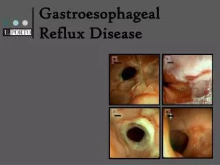
Anatomic radiographic landmarks of the lower esophageal sphincter (LES).
Gastroesophageal Reflux Disease (GERD) • Gastroesophageal reflux is a normal physiologic phenomenon experienced intermittently by most people, particularly after a meal. • Gastroesophageal reflux disease (GERD) occurs when the amount of gastric juice that refluxes into the esophagus exceeds the normal limit, causing symptoms with or without associated esophageal mucosal injury.
Definition • American College of Gastroenterology (ACG) • Symptoms OR mucosal damage produced by the abnormal reflux of gastric contents into the esophagus • Often chronic and relapsing • May see complications of GERD in patients who lack typical symptoms
Physiologic vs Pathologic • Physiologic GER • Postprandial • Short lived • Asymptomatic • No nocturnal sx • Pathologic GERD • Symptoms • Mucosal injury • Nocturnal sx
GERD Pathophysiology • Abnormal lower esophageal sphincter • or • Increase abdominal pressure
GERD Pathophysiology • Abnormal lower esophageal sphincter • Functional (frequent transient LES relaxation) • Mechanical (hypotensive LES) • Foods (eg, coffee, alcohol), • Medications (eg, calcium channel blockers), • Location .......... hiatal hernia • or B. Increase abdominal pressure The most common cause of (GERD). decrease the pressure of the LES. obesity Pregnancy increased gastric volume
Pathophysiology • Primary barrier to gastroesophageal reflux is the lower esophageal sphincter • LES normally works in conjunction with the diaphragm • If barrier disrupted, acid goes from stomach to esophagus
Summary of Pathogenesis of GERD • impaired lower esophageal sphincter-low pressures or frequent transient lower esophageal sphincter relaxation • hypersecretion of acid • decreased acid clearance resulting from impaired peristalsis or abnormal saliva production • delayed gastric emptying or duodenogastric reflux of bile • salts and pancreatic enzymes.
Clinical Manisfestations • Most common symptoms • Heartburn—retrosternal burning discomfort • Regurgitation—effortless return of gastric contents into the pharynx without nausea, retching, or abdominal contractions Atypical symptoms….coughing, chest pain, and wheezing.
Extraesophageal manifestations of GERD • Otolaryngeal: • hoarsness/laryngitis • Ch. Sore throat • Other: • Noncardial chest pain
Diagnostic Evaluation • If classic symptoms of heartburn and regurgitation exist in the absence of “alarm symptoms” the diagnosis of GERD can be made clinically and treatment can be initiated
Esophagogastrodudenoscopy • Endoscopy (with biopsy if needed) • In patients with alarm signs/symptoms • Those who fail a medication trial • Those who require long-term tx • The procedure lacks sensitivity for identifying pathologic reflux • Absence of endoscopic features does not exclude a GERD diagnosis • Allows for detection, and management of esophageal injury or complications of GERD
pH • 24-hour pH monitoring • Accepted standard for establishing or excluding presence of GERD for those patients who do not have mucosal changes • Trans-nasal catheter or a wireless, capsule shaped device
Complications • Erosive esophagitis • Stricture • Barrett’s esophagus
Complications • Erosive esophagitis • Responsible for 40-60% of GERD symptoms • Severity of symptoms often fail to match severity of erosive esophagitis
Esophagitis Eosinophils and neutrophils Elongation of lamina propria papillae basal zone hyperplasia,
Complications • Esophageal stricture • Result of healing of erosive esophagitis • May need dilation
Complications • Barrett’s Esophagus • Intestinal metaplasia of the esophagus • Associated with the development of adenocarcinoma 8-15%
Pathophysiology of Barrett’s Esophagus • Acid damages lining of esophagus and causes chronic esophagitis • Damaged area tries to heal in a metaplastic process and damaged squamous cells are replaced by metaplastic columnar cells defined by the presence of goblet cells (intestinal metaplasia) • This specialized intestinal metaplasia can progress to dysplasia and adenocarcinoma • Many patients with Barrett’s are asymptomatic
Complications dysplasia Barrett’s esophagus adenocarcinoma
The risk of cancer in Barrett's esophagus is estimated to be 40 to 100 times • Endoscopic surveillance is recommended for all patients with Barrett's esophagus. Endoscopy is performed every 2 years, and biopsies are taken from the area of abnormal mucosa. • If the biopsies reveal low-grade dysplasia, then the frequency of endoscopies is increased.
If high-grade dysplastic changes are seen and confirmed by a second pathologist, then the risk of subsequent adenocarcinoma is greater than 25%, and surgical resection should be considered.
Treatment • H 2 receptor Blockers • Proton pump inhibitors Antireflux surgery