Download
1 / 17
250 likes | 820 Views
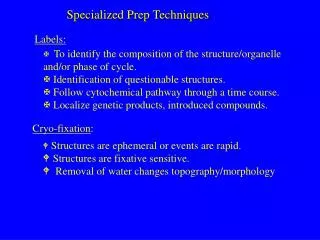
Specialized imaging techniques. Marilyn Rose RT, RDMS. Outline. Diagnostic Imaging Modalities Mammography- Mammo Ultrasound- US Computed tomography- CT Magnetic Resonance Imaging- MRI Nuclear Medicine Single- Photon Emission- CT- SPECT Positron Emission Tomography-PET Fusion Imaging.

E N D
Specialized imaging techniques Marilyn Rose RT, RDMS
Outline • Diagnostic Imaging Modalities • Mammography- Mammo • Ultrasound- US • Computed tomography- CT • Magnetic Resonance Imaging- MRI • Nuclear Medicine • Single- Photon Emission- CT- SPECT • Positron Emission Tomography-PET • Fusion Imaging “ As the world of technology advances, medical imaging modalities have become more technical. This change requires the radiographer to have a broader and more specific skill set to produce quality images. “ Eisenberg-Johnson 5th edition 2012
Mammography • FFDM- full-field digital mammo- most common • Faster with a lower dose of raditation • Screening- 2 images of each breast • Craniocaudal and mediolateral oblique projections • Diagnostic- 2 images plus 90 degree mediolateral projection • Ultrasound supplements mammo images • Lesion is cystic or solid
Ultrasound- US • Cross sectional imaging- • low cost, noninvasive, differentiate cystic, solid and complex tissue. • High frequency sound waves -stimulation of piezoelectric crystals. • Sound waves pass though the body- intensity reduced depending on acoustic properties • crystals in the transducers- send signals / receives echoes reflected back- recording changes in pitch and direction. • The monitor displays both the intensity level of echoes and position in the body. • Color display is for motion- blood flow- toward or away from the transducer • Anechoic- echo free- transmit sound easily- dark (black) • hyper/hypoechoic- solid structures- compare echo intensities with adjacent structures. • Isoechoic- two structures same echogenicity (liver= isoechoic to spleen)
Advantages of US • Safety! Modality for pregnant women and children. • Fetal age, placenta location, congenital anomalies, complications of pregnancy, intraperitoneal/ retroperitoneal structures, abdominal/ pelvic pathologies, obstruction of biliary and urinary tracts. • Pelvic US of prostate (transrectal) -staging of neoplasms • Pelvic US –ovarian/ uterine (transvaginal) • Imaging guide for bx, aspiration, drain placement • MSK- soft tissue mass, joints, fluid • QUALITY OF SCAN IS OPERATOR DEPENDENT.
US vascular Doppler – used to assess patency of major blood vessels- obstruction, stenosis, blood clot, plaque/ emboli. Duplex system- real time imaging, with Doppler- demonstrates motion, direction and velocity.
Computed tomography- CT • Cross sectional tomographic images- scanning a slice of tissue from multiple angles with a narrow x-ray beam then calculating the linear attenuation coefficient. • Displays CT reconstruction as a gray scale image • Sensitive to 1% differences in tissue density • CT number- Hounsfield= attenuation of specific tissue relative to water • Water= CT number 0- gray • Bone= CT number 1000- white • Air= CT number -1000- black • (fat numbers are <0 and soft tissues >0)
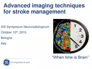
CT images • Window and level- midpoint of center of total densities- demonstrate parts of anatomy- lung, liver, bone. • IV contrast- • differentiation of vascular from nonvascular structures. • Differences in time of enhancement can detect neoplastic or infectious processes. • fat is a natural contrast- loss of fat plane suggests tumor extension • Abdominal studies- dilute oral contrast- 1-3% Barium is given to demonstrate GI tract from abdominal organs. CT image (window level = −500 HU,
Ct advancements • Conventional CT- section thickness of 5-10 mm • Multiple single scans • High-resolution- 1.5- 2 mm- lung anatomy • Spiral or Helical- CT scans as patient moves through gantry- faster scanning w/o respiratory motion- data can be reformatted. • May need 3 phase scanning with arterial, capillary/venous and excretory phase. Allows bolus to be distributed for better visualization. • 6th generation- multidetectors- 8-64 slices per rotation, pitch (table move) and variable rotation speed of the tube and instant 3D images- allow for advances in CT angio.
Magnetic Resonance Imaging-MRI • Modality of choice for CNS, spine and MSK. • fine tune questions of abdomen/pelvis • Induce hydrogen atoms (protons) to alternate between high and low energy states- absorbing and releasing energy. • Absorption is accomplished by placing the part of interest into a strong magnetic field and directing RF pulse • Protons absorb and move into high E • After pulse is turned off- transfer E and move back to low E= relaxation (T1 and T2) • The receiver coil placed on part of interest calculated the time it takes for relaxation and info is transferred to the computer as a gray scale image.
Mri image • MRI technologist selects a pulse sequence- scan parameter- with RF pulses and their timings- • varying TE and TR produces a weighted image- proton density. • Demonstrates differences in normal and abnormal tissue • T1 weighted • high signal intensity- bright- fat, sub-acute hemorrhage and gad • low signal intensity - dark- water, CSF • T2 weighted (opposite) • High signal- bright- water • Low signal- dark- muscle, soft tissue/ fat • No signal- very dark- cortical bone, calcium, air and fast flowing blood.
Mri advantages/disadvantages • Excellent spatial resolution of soft tissue • MRI angio- visible without contrast • rival conventional angio and soon replace for diagnosis of vascular disease. • Fat suppression. • Lack specificity in abnormal tissue- w/out contrast. • Long scanning time- image degradation- pt motion • Contraindicated for pacemaker, stent, clips • Cannot treat vascular disease at time of exam
New developments in mri • MRS- (mr spectroscopy) - chemical composition of tissue- benign vs malignant- breast/ prostate. • Diffusion imaging- movement of water molecules- changes with pathology- liver lesions, compression fx • Perfusion imaging- gad- measures transit time of blood – old vs new infarctions or ischemic regions • MRI can use fat suppression- soft tissue of neck, abd • Functional MRI (fMRI)-maps regions of the brain corresponding to functions- motor, sensory, memory, vision, language- patient performs a function and the area of brain being used show increase in blood flow.
Nuclear medicine • Patient ingests- iv or im- radiopharmaceutical that emits radiation from within. • no side effects like contrast reactions in CT • Amount of ionizing radiation in nuc med may equate to 25-1000 radiographs! • The gamma camera detects ionizing rad emitted from patient- interaction produces scintillation and is converted to digital signal on the monitor. • Can see changes in physiology earlier than in radiographic images. • Excellent for organ physiology- lack atomic information
Single-Photon Emission CT- SPECT • Nuc med imaging- gamma camera rotates independently around the patient. • 3D images- by acquisition of projections 180-360 degrees around the patient- the patient emits the signal and the detector rotates. • Multiple images can be sewn together to create a single whole body image. • Costs ½ the price of PET- but small region of interest • Coronary artery disease, ventricular d/o, infection, tumor, stroke, seizure, traumatic brain
Positron Emission Tomography-PET • Radiopharmaceutical s different because it decays by positron emission. • The positron interacts with an electron causing annihilation- producing two high-energy photons 180 degrees from each other. • The time it takes for each ray to reach the camera – determines the annihilation location which creates the image. • The body/ organ metabolism illustrates the biochemistry of the tissue- tissue viability
Fusion Imaging • Integrated imaging- accomplished with software to overlay/ fuse multidimensional computed data from MRI, CT , nuc med, SPECT, or PET into one single image. • Direct fusion equipment- hybrid technology • Exams are simultaneous and fusion software puts the data together • Lymph node staging