Download

1 / 18
250 likes | 535 Views
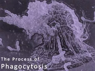
Phagocytosis. PMNLs Plasma cells Macrophage lymphocyte inflammation: seen here are mainly neutrophils , but there are also plasma cells, lymphocytes, and macrophages. GRANULOMA. Cont. Inflammation. Granuloma :.

E N D
PMNLsPlasma cells Macrophage lymphocyte inflammation: seen here are mainly neutrophils, but there are also plasma cells, lymphocytes, and macrophages
GRANULOMA Cont. Inflammation
Granuloma: -It’s a type of chronic specific inflammation, characterized by accumulation of tumor like masses. -types: 1-infective, 2-non-infective.
Granuloma: The single tubercle composed of : 1-central necrotic tissue, 2-epithellioid cells, 3-lymphocytes and plasma cells, 4-langhans cell .
Tuberculosis -Tuberculosis , MTB or TB (short for tubercles bacillus) is infectious disease caused by Mycobacterium tuberculosis -It is usually attacks the lungs but can also affect other parts of the body. - It is spread through the air when people who have active MTB infection cough, sneeze, or spit
1- Early T.B in lymph node: -Pale pink rounded tubercles among blue lymphoid tissue. -the tubercle is formed of : epithelioid cells, langhansgient cells &lymphocytes.
2- Caseating T.B in LN -Caseous is a form of biological tissue death. Necrosis (death) Tuberculosis - it is characterized by acellular pink areas of necrosis surrounded by a granulomatous inflammatory process
3- Chronic fibrocasious T.B in lung: -large irregular areas of caseation appearing homogenous pink surrounded by tubercles reaction an fibrous tissue.
Bilharziasis -It is a parasite infection by a trematode worm acquired from infested water. Also known as (schistosomiasis). - it is can produce in liver, bladder, and gastrointestinal problems
4- Bilharziasis in rectum: Bilharzia ova deposited mainly in the submucosa -ova appear rounded or oval and yllowishrefractileshell,degenerated ova are pink , while clascified ova are blue. -ova are surrounded by macrophages ,plasma cells, esinophils,lymphocytes & foreign body giant cells.
5- Bilharziasis in Urinary Bladder: -Bilharzia ova deposited mainly in the submucosa with less degree in other layers . -The ova surrounded by bilharzialrxn
6- Bilharziasis in liver: Bilharzias ova deposited in the portal tract. The ova surrounded by bilharzialrxn formed capilleries & new bile ducts.