Download
1 / 30
360 likes | 890 Views
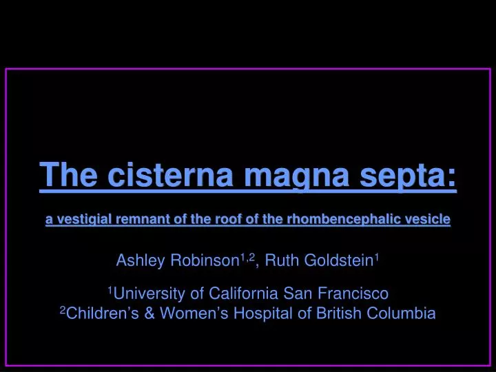
The cisterna magna septa: a vestigial remnant of the roof of the rhombencephalic vesicle. Ashley Robinson 1,2 , Ruth Goldstein 1 1 University of California San Francisco 2 Children’s & Women’s Hospital of British Columbia. The cisterna magna septa Introduction. The phrase:

E N D
The cisterna magna septa:a vestigial remnant of the roof of the rhombencephalic vesicle Ashley Robinson1,2, Ruth Goldstein1 1University of California San Francisco 2Children’s & Women’s Hospital of British Columbia
The cisterna magna septa Introduction • The phrase: “Posterior fossa cyst communicating with fourth ventricle” Dandy-Walker malformation however it is actually a description of normal developmental anatomy
The cisterna magna septa Introduction • The cisterna magna septa: • are seen in up to 92% of fetuses in 2nd and 3rd trimesters • usually straight, arising at the cerebellovermian junction • course directly to the occipital bone axial
Typical appearances 22 weeks • Septa become more difficult to discern later in gestation • even in the same fetus • but the posterior fossa structures develop normally
Typical appearances Same fetus at 28 weeks Septa less visible
Typical appearances Same fetus at 35 weeks Septa not visible Structures appear to have developed normally
Typical appearances28 weeks - neonatal Above vallecula 4th ventricle walls continuous with septa via vallecula 4th ventricle walls separate from septa
Typical appearances28 weeks At vallecula 4th ventricle walls continuous with septa via vallecula CSF space enclosed between septa is in direct continuity with CSF space of the fourth ventricle
Variable appearances28 weeks - neonatal axial • The falx cerebelli is visible as a midline septum superiorly in the posterior fossa
Variable appearances27 weeks - neonatal • Sometimes the septa • and falx cerebelli • can be seen as three septa on the same scan plane
Typical appearances20 weeks coronal • space between the septa is always completely anechoic • space outside of the septa is usually slightly echogenic • especially at earlier gestations
Embryology of the dura epidural conjunctive tissue containing rich vascular plexi meninx primitiva developing cerebrum
Embryology of the dura as hemispheres overgrow midline structures dura become apposed the vascular plexi coalesce and meninx primitiva cavitates
Embryology of the dura where dural leaves don’t fuse sinuses form superior sagittal inferior sagittal
Colour Doppler 21 weeks If the cisterna magna septa were dural leaves there would vascular spaces between them flow in occipital sinus no flow between septa
The cisterna magna septaDiscussion • We therefore don’t agree that the septa represent • dural folds - inferior attachments of falx cerebelli • Pretorius DH, Kallman CE, Grafe MR et al. Linear echoes in the fetal cisterna magna. J Ultrasound Med. 1992 Apr;11(4): 125-8 • the Torcula Herophili • Pilu G, Romero R, De Palma L, et al. Ultrasound investigation of the posterior fossa in the fetus. Am J Perinatol. 1987 Apr;4(2): 155-9 • the straight sinus • Mahony BS, Callen PW, Filly RA, et al. The fetal cisterna magna. Radiology. 1984 Dec;153(3): 773-6
The cisterna magna septaDiscussion • We also don’t agree that the septa represent bridging arachnoid septations • Knutzon RK, McGahan JP, Salamat MS, et al. Fetal cisterna magna septa: a normal anatomic finding. Radiology. 1991 Sep;180(3): 799-801 • they are thicker than other pia-arachnoid septations seen in the subarachnoid space • We believe that the cisterna magna septa represent the walls of Blake’s pouch • Blake JA. The roof and lateral recesses of the fourth ventricle considered morphologically and embyrologically. J Comp Neurol 1900;10: 79-108
Normal embryologyrhombencephalon sagittal focal dilatation of the central canal of the neural tube rhombencephalic vesicle
Normal embryologyrhombencephalon sagittal Dorsal pontine flexure Transverse crease Anterior membranous area (AMA) Posterior membranous area (PMA)
Normal embryologyrhombencephalon sagittal AMA develops into vermis Choroid plexus forms in crease Cavitation starts in meninx primitiva
Normal embryologyrhombencephalon sagittal PMA evaginates = ependyma-lined diverticulum into the meninx primitiva Blake’s pouch Further cavitation in meninx primitiva
Normal embryologyrhombencephalon sagittal Multiple pia-arachnoid trabeculations Blake’s pouch fenestrates variably down to obex neck of Blake’s pouch = foramen of Magendie
Normal embryologyrhombencephalon fluid more echogenic trabeculated by pia-arachnoid septations axial As Blake’s pouch expands walls are visible as cisterna magna septa fluid anechoic intra-axial
Lateral displacement of the septa 24 weeks neonatal • The septa are sometimes deviated laterally giving impression of a cyst • Fenestration of Blakes’ pouch variable in timing • Prior to this, increased intra-axial CSF pressure may lead to the transient enlargement of 4th ventricle that has been demonstrated at 14-16 weeks, resolving by 22-23 weeks gestation • Bronshtein M, Zimmer EZ, Blazer S. Isolated large fourth ventricle in early pregnancy--a possible benign transient phenomenon. Prenat Diagn. 1998 Oct;18(10): 997-1000 • Knutzon RK, McGahan JP, Salamat MS, et al. Fetal cisterna magna septa: a normal anatomic finding. Radiology. 1991 Sep;180(3): 799-801
Lateral displacement of the septa 24 weeks neonatal • outward bowing of the cisterna magna septa may be due to this delay in fenestration
Conclusions • Our findings support current theories that • Blake’s pouch cyst • Dandy-Walker continuum • Mega cisterna magna • are a single spectrum of developmental abnormalities of roof of the rhombencephalic vesicle
Conclusions • differences depend on: • degree of dilatation of Blake’s pouch • degree & timing of fenestration of the 4th ventricular outlet foramina • degree of vermian hypoplasia
Elevation of the vermis • probably simply due to an expanded Blake’s pouch • may lead to false-positive diagnosis of vermian hypoplasia
Conclusions • The cisterna magna septa • represent the walls of Blake’s pouch, a normal dorsal expansion of the roof of the rhombencephalic vesicle into the cisterna magna • may be potential new marker for normal development of roof of the rhombencephalon
Thankyou for your attention Please also see our poster OP 03.17