Download
1 / 62
620 likes | 629 Views
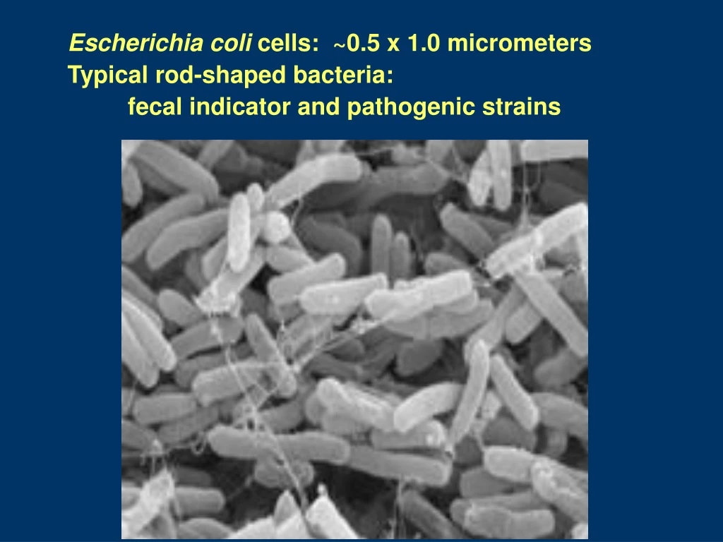
Escherichia coli cells: ~0.5 x 1.0 micrometers Typical rod-shaped bacteria: fecal indicator and pathogenic strains. E. coli Genetics and Serology. Genetics: Single, circular DNA molecule, ~4 x 10 6 base pairs Molecular weight of 4 x 10 9 Total length of about 1.4mm. Serology:

E N D
Escherichia coli cells: ~0.5 x 1.0 micrometers Typical rod-shaped bacteria: fecal indicator and pathogenic strains
E. coli Genetics and Serology Genetics: • Single, circular DNA molecule, ~4 x 10⁶ base pairs • Molecular weight of 4 x 10⁹ • Total length of about 1.4mm. Serology: • E. coli can be subdivided by somatic (cell-wall) or O antigens and flagellar or H antigens. • >160 recognized O types and 55 recognized H types • over 8000 possible OH serotypes. • also capsular (K) and fimbrial antigens.
Virulence Properties of E. coli Enterotoxins: • at least two types: Heat Stable (ST) and Heat Labile (LT) • Verotoxins or Shiga-like toxins (interchangeable terms): • Verotoxin term is based on the reactions of toxins on Vero cells • VT1 (SLT I): similar to Siga-toxin (produced by some strains of Shigella dysenteriae) • VT2 (SLT II) which is only about 50% realted Shiga toxin. • Other Toxins: • Cytolethal distending toxin (CLDT), VirCytotoxin, Cytotoxic necrotising factors (CNF), a possible Enteropathogenic E. coli EPEC) enterotoxin and a possible E. coli Sudden Infant Death Syndrome (SIDS)-toxin. • Haemolysins: • extracellular haemolysin known as alpha-haemolysin (many strains) • cell-associated haemolysin, beta-haemolysin, (some strains) • enterohaemolysin: extracellular; Enterohaemorrhagic E. coli (EHEC)
Pathogenic E. coli Enteric Infections: • Enteroadherent E. coli (EAEC) • Enteroaggregative E. coli (EAggEC) • Enterohaemorrhagic E. coli (EHEC) • Enteroinvasive E.coli (EIEC) • Enteropathogenic E. coli (EPEC) • Enterotoxigenic E. coli (ETEC) Extraintestinal Infections: • Uropathogenic E. coli (UPEC): urinary tract infections • Neonatal Menigitis E. coli (NMEC).
Virulence Properties of E. coli • Fimbriae: CFAI/CFAII, Type 1 fimbriae, P fimbriae, S fimbriae • most important: K88, K99 and CFA fimbriae associated with enterotoxigenic E. coli (ETEC). They have differing species specificities. • The p-fimbriae: associated with urinary tract pathogens. • E. coli also produce common fimbriae not associated with virulence. • Adhesins: • Intimin: non-fimbrial adhesin; causes the intimate association with target cells in enteropathogenic and enterohaemorrhagic E. coli . • Associated with the 'attachment and effacement' phenomenon • Causes destruction of the intestinal surface cells. • Other outer membrane proteins can act as adhesins.
Shigella and Shigellosis • Fecal-oral transmission • person-to-person, fomites, food, water, ect. • Waterborne and water-washed • Reservoirs: humans and primates • Infectious dose: low; as few as 10 cells to infect • Incubation period: 1 to 7 days; typically, 1-3 days • Duration of illness: • untreated: severe symptoms for about two weeks • Antibiotic treatment shortens illness and prevent spread to others
Shigellosis - Epidemiology • Four species of Shigella: flexneri, sonnei, dysenteriae, boydii • Major public health problem in many developing countries • causes about 5 to I0% of childhood diarrhoea • up to 25% of all diarrhea-related deaths can be associated with Shigella Developing countries: • Sh. flexneri is endemic (always present) in most communities • Sh. dysenteriae type 1 often occurs in an epidemic pattern • organism can be absent for a number of years, then reappear and infect a large proportion of the population. • These two species of Shigella generally produce the most severe illness. Developed countries: • Sh. sonnei is the most common and is the least virulent • Sh. boydii causes disease of intermediate severity • is least common, except in the Indian sub-continent.
Salmonella and Salmonellosis • Belong to Enterobacteriaceae family • Gram-negative bacilli; facultative and flagellated (motile). • 3 major antigens: • "H" or flagellar antigen (phase 1 & 2) • "O" or somatic antigen (part of the LPS moiety) • "Vi" or capsular antigen (called "K" in other Enterobacteriaceae). • Posess LPS endotoxin characteristic of Gram-negative bacteria • composed of an "O” polysaccharide ("O" antigen) • "R" core • endotoxic inner "Lipid A". • Endotoxins evoke fever and can activate complement, kinin and clotting factors.
Typhoid fever: (S. typhi and S. paratyphi): Systemic Infection • Fecal-oral transmission • Systemic infection: • Macrophages, reticuloendothelial system (esp. liver, spleen and bone marrow), gallbladder and intestines as major sites of damage • 1.5‑2 week incubation period • Symptoms: fever, headache, malaise, anorexia, then bloody diarrhea • Mortality rate 10%, if untreated • Carrier state possible • "Typhoid Mary”: infamous food handler; infected hundreds • Fecally shed at billions/gram by ill persons and carriers
Yersinia pestis • Gram Stain: • Small, gram-negative bipolar-stained coccobacilli • Wayson Stain: • Pink-blue cells with a closed safety pin look
Yersinia pestis: Plague • U.S. averages 13 cases/yr (10 in 1998) • 30% of cases are in Native Americans in the Southwest. 15% case fatality rate • Most cases occur in summer
Plague Epidemiology • Three Clinical Types: • bubonic (infected lymph nodes) • septicemic (blood-borne organisms) • pneumonic (transmissible by aerosol; deadliest)
Legionella spp. • Gram-negative • Aerobic • Non-sporing • Encapsulated • ~46 species, 68 serogroups • Ubiquitous aquatic organism • Thrives in warm environments (32C-45C)
Legionella: Legionellosis and Pontiac Fever Reservoirs and amplifiers: • Hot water systems • circulating water ventilation systems (cooling towers) • Plumbing (e.g., shower heads). • Hot tubs, whirlpools, etc. • Produce fresheners Cleveland Auto plant outbreak, March, 2001: • Plant cooling tower is considered a possible source of the outbreak. • But, more than 100 other internal water sources -- favorite breeding grounds for the Legionella bacteria -- were also under investigation….
Legionnaire’s Disease and Pontiac Fever Legionnaire's disease: • Bacterial pneumonia caused by Legionella pneumophila. • A type of pneumonia that affects the lungs and may also affect the stomach and intestines, kidneys, and central nervous system. • Incubation period: 2-10 days after exposure • Frequently requires hospitalization • Aerosol exposure from contaminated cooling towers, evaporative condensers, whirlpools, shower heads, faucets, & hot water tanks. Pontiac fever: also caused by Legionella. • A "flu-like" illness with fever, chills, headache, myalgia (pain in the muscles), cough, nausea, and breathlessness. • Pneumonia does not occur. • Usually lasts 2-5 days. • Same sources as for Legionnaires' disease
Kingdom Fungi • Major role in recycling of organic matter-”Saprophytes” or “Saprobes” • Eukaryotic, unicellular or multicellular structures with rigid chitinous cell wall • No chlorophyll • Asexual and sexual reproduction • Anamorph (mitosporic?) or Telomorph (meiosporic?) • Homothallic (resources to reproduce asexually in single organism) or heterothallic (sexes)
Yeasts – single-celled organisms; cell wall – chitin; typical eukaryotic cell; reproduce by budding; aerobic to anaerobic Molds – hypha (hyphae) – long thin filaments; aggregated hyphae form the mycelium; cell wall of chitin Dimorphic fungi – can exist as either mold form or yeast form; may be temperature dependent e.g. room temp – mold body temp – yeast (Penicillium marneffi, Coccidiodes immitis)
Fungal Structures • Hyphae • Spores • Conidia (asexual spores)
Medically Relevant Fungi • Four Major Phyla: • Ascomycota (ascospores) • Basidiomycota (basidiospores) • Zygomycota (zygospores) • Microsporidia • Fungi Imperfecti (no longer considered correct) • Deuteromycota Dikarya
Spores • Basidiomycota – mushrooms, smuts, rusts, e.g. Cryptococcus • Ascomycota – sac fungi, yeasts, morels, truffles, e.g. Pennicillium, Aspergillus • Zygomycota – e.g. Mucor
Ascomycetes – sac fungi red, brown and blue-green fungi associated with food spoilage plant diseases chestnut blight and Dutch elm disease edible morels and truffles examples:Morchella esculentacommon morel Claviceps purpurea – parasite of rye and other grasses disease called ergot ergotism in humans – poisoning in humans St. Anthony’s fire – killed thousands in Middle Ages psychotic delusions, nervous spasms, convulsions ergot alkaloids – LSD
Basidiomycetes – club fungi smuts, rusts, shelf fungi, puffballs, toadstools, mushrooms basidium – club-shaped structure on which spores are produced basidiocarp – mushroom most are saprophytes
Basidiomycetes – club fungi smuts, rusts, shelf fungi, puffballs, toadstools, mushrooms basidium – club-shaped structure on which spores are produced basidiocarp – mushroom most are saprophytes
Basidiomycetes – club fungi examples: smuts & rusts – crop damage in millions Agicarium campestris – multimillion-dollar business Amanita phalloides – toxic two toxins – phalloidin and α-amanitin phalloidin – liver toxinα-amanitin – lining of the intestinal tract Cryptococcus neoformans – cryptococcosis lungs and central nervous system
Zygomycetes most soil organisms a few pathogens of plants, insects, animals form thick-walled zygospores sexual reproduction – fusion of + and - strains examples: bread mold Rhizopus; black spores Mucor – production of sofu from soybeans some pathogens
Fungal Disease • Infection (mycosis) • Allergy • Hypersensitivity Pneumonitis (HP) and Organic Dust Toxic Syndrome (ODST) • Toxicosis • VOCs???
Fungal Infections • Mycosis: any fungal Infection • Systemic mycoses: fungal infection deep inside body. Can result from inhalation of spores. Ex: Valley fever (coccidioidomycosis) • Subcutaneous mycoses: infection beneath skin as a result of puncture
Fungal Infections, cont. • Cutaneous mycoses: infections of epidermis, hair, and nails. Direct contact with infected person, animal, hair, or cells is required. • Superficial mycoses: infections of hair shaft and surface (dead) epidermal cells • Opportunistic: compromised host
MEDICALLY IMPORTANT FUNGI Opportunistic - compromised host Candidiasis – vaginal infections 10% of blood infections in hospital infections thrush – oral infection
Fungal Toxins • Most fungal toxins produced by: • Aspergillus spp. • Penicillium spp. • Fusarium spp. • Alternaria spp. • Rhizopus • Mucor
Aflatoxins Fumonisins Ochratoxins Trichothicenes Zearalenone and Zearalenoles Tenuazonic Patulin Xanthomegnin and Viomellein Nephrotoxic glycopeptides Sterigmatocystin Fusarochromanone Ergot alkaloids Citrinin 3-Nitropropionic acid Roquefortine C Secalonic acid D Verrucosidin Important Mycotoxins
Dermatophytosis (Ringworm and Tinea) Dermatophytes which have the ability to utilize keratin as a nutrient source. The disease process in dermatophytosis is unique for two reasons: No living tissue is invaded; the keratinised stratum corneum is simply colonized. - the presence of the fungus and its metabolic products usually induces an allergic and inflammatory eczematous response in the host. 2. The dermatophytes are the only fungi that have evolved a dependency on human or animal infection for the survival and dissemination of their species. Tinea is general term for skin mycoses; not taxonomical; Charaterized by location – e.g. Tinea pedis (athletes foot), Tinea cruris (Jock itch), Tinea corporis (ringworm Can be caused by variety of fungi – e. g. Trichopyton spp., Microsporum spp.
Tinea nigra. Tinea nigra is a superficial fungal infection of skin characterized by brown to black macules, which usually occur on the palmar aspects of hands and occasionally the plantar and other surfaces of the skin. Lesions are non-inflammatory and non-scaling. Distribution: World-wide, but more common in tropical regions of Central and South America, Africa, South-East Asia and Australia. Aetiological Agent:Exophiala werneckii a common saprophytic fungus believed to occur in soil, compost, humus and on wood in humid tropical and sub-tropical regions. Familial spread of infection reported.
Tinea Versicolor Pityriasis; A chronic, superficial fungal disease of the skin characterized by well-demarcated white, pink, fawn, or brownish lesions, often coalescing, and covered with thin furfuraceous scales. Lesions occur on the trunk, shoulders and arms, rarely on the neck and face, and fluoresce a pale greenish colour under Wood's ultra-violet light. Young adults are affected most often, but the disease may occur in childhood and old age. Distribution: World-wide but more common in tropical than temperate climates. Aetiological Agent:Malassezia furfur a lipophilic yeast forming part of the normal flora of human skin.
Histoplasmosis An intracellular mycotic infection of the reticuloendothelial system caused by the inhalation of the fungus. Approximately 95% of cases of histoplasmosis are subclinical. Five percent of the cases have chronic progressive lung disease, chronic cutaneous or systemic disease or an acute fulminating fatal systemic disease. All stages of this disease may mimic tuberculosis. Distribution: World-wide, especially U.S.A. Sporadic cases do occur in Australia. Aetiological Agent:Histoplasma capsulatum, especially from soil enriched with excreta from chicken, starlings and bats.
Coccidioidomycosis Initially, a respiratory infection, resulting from the inhalation of conidia, that typically resolves rapidly leaving the patient with a strong specific immunity to re-infection. However, in some individuals the disease may progress to a chronic pulmonary condition or as a systemic disease involving the meninges, bones, joints and subcutaneous and cutaneous tissues. Distribution: Endemic in south-western U.S.A., northern Mexico and various centres in South America. Aetiological Agent:Coccidioides immitis, a soil inhabiting fungus.
Subcutaneous zygomycosis (Mucormycosis) • Usually the result of a "barrier break" or traumatic implantation of fungal elements. • Lesions vary considerably in morphology but include plaques, pustules, ulcerations, deep abscesses and ragged necrotic patches. • Most heal with little treatment (debridement and amphotericin B) and they are not usually associated with dissemination. • Distribution: World-wide. • Aetiological Agents: Cosmopolitan members of the Mucorales including species of Rhizopus, Mucor, Rhizomucor, Absidia, Cunninghamella, Saksenaea and Mortierella.