Download
1 / 7
70 likes | 75 Views
Easton et al. 1. αCaMKII autophosphorylation controls the establishment of alcohol drinking behaviour Supplement information. Methods and Materials Animals

E N D
Easton et al. 1 αCaMKII autophosphorylation controls the establishment of alcohol drinking behaviour Supplement information Methods and Materials Animals Mutants were generated using a gene-targeting strategy which utilizes a replacement vector containing the point mutation and a neo gene flanked by loxP sites; after homologous recombination the neo gene was removed by Cre recombination [1]. R1 ES cells (F1 between 129/Sv and 129/Sv-CP) were used. The mice were subsequently backcrossed for at least eight generations into C57BL/6J and then crossed once with 129S2/SvHsd mice and kept in this mixed background by interbreeding (mice had gone through more than four rounds of interbreeding). Mice were obtained from interbreeding of heterozygotes and genotypes were determined by PCR with tail biopsies as described [2]. In the homozygous mutants (MT) the autophosphorylation of αCaMKII is prevented due to the insertion of a missense mutation (threonine-286 changed to alanine; T286A) within the autoinhibitory domain [1]. Animals were housed in Tecniplast cages (32cm x 16cm x 14cm), using Litaspen sawdust and nesting materials, (Sizzlenest, Datsand, Manchester UK). Determination of blood alcohol levels Briefly, after predilution of the samples to obtain appropriate volume, if necessary, the samples were assayed with Integra 400 Plus (Roche) analyzer. In the presence of nicotinamide adenine dinucleotid (NAD+), alcohol dehydrogenase oxidizes ethanol to acetaldehyde. Simultaneously generated NADH, which is directly proportional to the ethanol concentration in the sample, is measured photometrically. In each analytical run, appropriate quality control samples were analyzed. In-vivo Microdialysis Surgery The mice were deeply anaesthetised with an intraperitoneal (i.p.) injection using a mixture of 4.12ml saline (NaCl), 0.38ml Ketaset (containing 100mg/ml Ketamine) and 0.5ml Domitor (containing 1mg/ml Medetomidine hydrochloride) administered at 0.1ml per 10g body weight. In addition 0.01ml Rimadyl (5mg/kg Carprofen) analgesia was given subcutaneously (s.c.). The animal was placed in a Kopf stereotaxic frame. Two guide cannulas (Microbiotech/se AB, Stockholm, Sweden) were aimed at the prefrontal cortex (PFC) (A: +1.9; L: ±0.8; V: -1.3 angle ±10º from midline) and the nucleus accumbens (NAcc) (A: +1.2; L: ±1.6; V: -4.3 angle ±10º from midline) using coordinates relative to bregma [8], and fixed in place using two anchor screws (stainless steel, d=1.4mm) and dental cement. Anaesthesia was reversed by administering a mixture of 3.9ml saline (NaCl) and 0.1ml Antisedan (containing 5mg/ml Atipamezole) at 0.08ml/ 10g body weight (s.c.) after approximately 45 minutes. Animals were kept warm and allowed to recover from the anaesthetic. Animals were then returned to their home cages and monitored daily, allowing at least 5 days for complete recovery. Procedure The microdialysis probes were connected to a microinfusion pump (CMA 400, Carnegie, Sweden) via a swivel mounted on a balanced arm above the chamber, and were perfused with artificial cerebrospinal fluid (aCSF) (containing Na+ 147 mmol, K+ 4 mmol, Ca2+ 2.2 mmol, Cl- 156 mmol, pH = 7.4) at room temperature [9]. The flow rate was set to 1.5μl/min and allowed to stabilise for at least two hours until a stable baseline was obtained. Samples were collected every 20 minutes into vials containing 2.73μl of antioxidant (0.1 M perchloric acid and 500 pg dihydroxybenzylamine (DHBA) as internal standard).
Easton et al. 2 HPLC-ED analysis: The column was an ET 125/2, Nucleosil 120-5, C-18 reversed phase column (Macherey–Nagel, Germany) perfused with a mobile phase composed of 75 mM NaH2PO4, 4 mM KCl, 20μM ethylenediamine tetraacetic acid (EDTA), 1.5 mM sodium dodecyl sulfate, 100 μl/l diethylamine, 12% methanol, and 12% acetonitrile adjusted to pH 6.0 using phosphoric acid. The electrochemical detector (Intro, Antec, The Netherlands) was set at 500 mV vs. an in situ Ag/AgCl (ISAAC) reference electrode (Antec, Leyden, Netherlands) at 30°C. This setup allows the simultaneous measurement of DA and 5-HT. The detection limit of the assay was 0.1pg for all neurotransmitters with a signal–noise ratio of 2:1. Neurochemical data were not corrected for recovery [3, 4]. c-Fos activation after acute and subchronic alcohol treatment Procedure Mice were culled under isoflurane narcosis and transcardially perfused with 0.1M PBS for 10 minutes and then fixed with 4% paraformaldehyde (PFA) solution for a further 10 min (flow rate 4ml/min). The brain was removed and left in 4% PFA solution overnight at 4°C. Brains were then transferred to a 30% saccharose solution and stored at 4°C until brains were fully submerged. Brains were then snap frozen in isopentane at -60°C and stored at -80°C until the whole brain was cut into 40 µm coronal sections by cryosectioning. All sections were collected and then stored at -20°C in an anti-freezing solution until processed for immunohistochemical staining. The floating coronal sections were incubated with an anti-c-Fos rabbit polyclonal antibody (1:30.000, Calbiochem, Germany) for 20 hours. c-Fos immunoreactive cells were visualized using a biotinylated donkey anti-rabbit secondary antibody (1:500, Santa Cruz, Germany) and the avidin-biotin complex (ABC-Elite kit rabbit, Vector Laboratories, Germany Stereological quantification The number of c-Fos-immunoreactive cells in the rostral and caudal ventral tegmental area (VTA) was determined using stereological quantification. The examined regions were defined according to the stereotaxic coordinates [5]. Stereological quantification of the c-Fos positive cells was carried out strictly blind to the experimental conditions with the optical fractionator estimating total numbers of c-Fos positive cells [6, 7]. After histological processing the sections had a mounted section thickness of 30 µm, a fixed distance of 2 µm and an optical dissector height of 26 µm, measured with an electronic microtator attached to the microscope. All counting procedures and measurements of reference volumes were conducted on a light microscope (Nikon Eclipse 80i) equipped with a semiautomatic stereology system (Stereoinvestigator, Version 8.27, MicroBrightField, Colchester, Vermont, USA). C-Fos-positive cells were counted within a 70x70 µm counting frame, which was spaced in a 90x90 µm counting grid. Positive cells were counted, if their nucleus came into focus. Positive cells, which intersected the uppermost focal plane (exclusion plane) or the lateral exclusion boundaries of the counting frame were not counted. The total counts of positive c-Fos cells were multiplied by the ratio of reference volume to sampling volume in order to obtain the estimated number of c-Fos-positive cells for each structure.
Easton et al. 3 c-Fos and GAD6t double labeling For immunofluorescence the floating coronal sections were incubated with an anti-c-Fos rabbit polyclonal (1:7000, Calbiochem, Germany) and GAD67 (1:300) goat polyclonal antibodies for 20 hours. c-Fos and GAD67 immunoreactive cells were visualized using fluorophore-coupled secondary antibodies: donkey anti-rabbit Alexa 488 (Jackson ImmunoResearch Laboratories, Inc., Germany) and donkey ant-goat Cy3 (Jackson ImmunoResearch Laboratories, Inc., Germany) in a dilution of 1:400. After 1 hour of incubation with secondary antibodies, sections were repeatedly washed in PBS, mounted, and coverslipped in Fluoromount™ Aqueous Mounting Medium (Sigma-Aldrich, Germany). Statistical Analysis Alcohol drinking: Drinking and taste preference data were analysed using two-way ANOVAs followed pre-planned comparisons using Fisher’s LSD with Bonferroni correction. Microdialysis: Baseline neurochemical and behavioural data were analysed using one way ANOVAs and pre-planned Fisher’s LSD tests. Planned t-test pair-wise comparisons were performed to determine differences between acute and subchronic time points within each genotype group. Alcohol induced neurochemical effects were expressed as a percentage of the mean of the three baseline samples which were taken as 100%. Data were compared using two-way ANOVAs for factor genotype (3) and time (12). To compare alcohol effects of certain time points pre-planned comparisons were performed using Fisher’s LSD tests. Planned t-test pair-wise comparisons were performed to determine differences between acute and subchronic time points within each genotype group. LORR: Data were analysed using two-way ANOVAs for factor genotype (3) and time (2). To compare alcohol effects across time, within genotype groups, pre-planned comparisons were performed using Fisher’s LSD tests. Blood alcohol levels: Data were analysed using two-way ANOVAs for factor genotype (3) and time (3). To compare alcohol effects across time and within genotype groups, pre-planned comparisons were performed using Fisher’s LSD tests. c-Fos expression: all data were analyzed by one-way ANOVAs followed by t-tests.
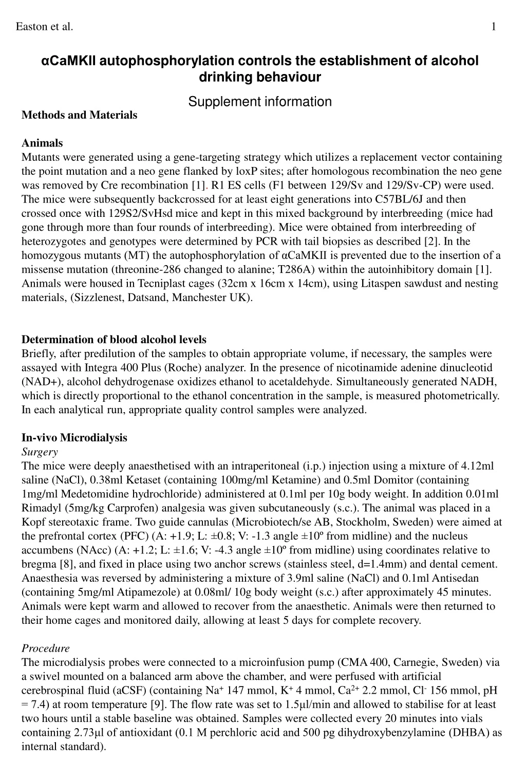
PFC Dopamine A). Acute B). Subchronic NAcc Serotonin C). D). Acute Subchronic Easton et al. 4 Supplement-Figure. 1. Microdialysis results are represented as percent of baseline. Graphs show extracellular dopamine (DA) levels in the prefrontal cortex (PFC) after acute (A) and subchronic alcohol treatments (B). Extracellular serotonin (5-HT) levels in the nucleus accumbens (NAcc) after acute (C) (two-way ANOVA, p>0.05; time: F11,308= 2.02, p=0.03)and subchronic (D) alcohol treatments. Pairwise comparisons revealed differences between WT and αCaMKIIT286A mice 20 min (LSD, p<0.05), 40 min (p=0.04), 80 min (p=0.02), and 140 min (p=0.009) after alcohol administration. Arrow indicates time of alcohol injection. Pre-planned comparison between groups at each time point, *p<0.05, #p<0.01 MT vs. WT.
A B GAD GAD C D c-Fos c-Fos E F merged merged Supplement-Figure. 2. GAD67 and c-Fos labelled cells of the caudal (A, C, E) and rostral ventral tegmental area (VTA, B, D, F) after alcohol (2 g/Kg, i.p.) treatment. C-Fos and GAD67 were determined 70 min after alcohol injection(bar=100 µm).
Easton et al. 5 Supplement-Table. 1 Haplotype based association tests of markers located in CAMK2A transcript region, using only haplotypes within blocks and with minimal frequency of 0.01 in whole sample.
Easton et al. 6 • 1. Giese, K.P., Fedorov, N.B., Filipkowski, R.K. & Silva, A.J. Autophosphorylation at Thr286 of the alpha calcium-calmodulin kinase II in LTP and learning. Science 279, 870-873 (1998). • Irvine, E.E., Vernon, J. & Giese, K.P. AlphaCaMKII autophosphorylation contributes to rapid learning but is not necessary for memory. Nat Neurosci 8, 411-412 (2005). • Pum, M., Carey, R.J., Huston, J.P. & Müller, C.P. Dissociating effects of cocaine and d-amphetamine on dopamine and serotonin in the perirhinal, entorhinal, and prefrontal cortex of freely moving rats. Psychopharmacology (Berl) 193, 375-390 (2007). • Pum, M.E., Huston, J.P., De Souza Silva, M.A. & Müller, C.P. Visual sensory-motor gating by serotonin activation in the medial prefrontal and occipital, but not in the rhinal, cortices in rats. Neuroscience 153, 361-372 (2008). • Franklin, K.B.J. & Paxinos, G. The mouse brain in stereotaxic coordinates (Academic Press, San Diego, 1997). • Gundersen, H.J. Stereology of arbitrary particles. A review of unbiased number and size estimators and the presentation of some new ones, in memory of William R. Thompson. J Microsc 143, 3-45 (1986). • Schmitz, C., et al. Use of cryostat sections from snap-frozen nervous tissue for combining stereological estimates with histological, cellular, or molecular analyses on adjacent sections. J Chem Neuroanat 20, 21-29 (2000). • Franklin, K.B.J. and G. Paxinos, The mouse brain in stereotaxic coordinates. 2nd ed1997, San Diego: Academic Press. • Lenz, B., C.P. Müller, C. Stoessel, W. Sperling, T. Biermann, T. Hillemacher, et al., Sex hormone activity in alcohol addiction: integrating organizational and activational effects. Prog Neurobiol, 2011. 96(1): p. 136-63.