Download
1 / 45
680 likes | 1.08k Views
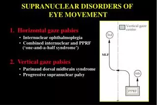
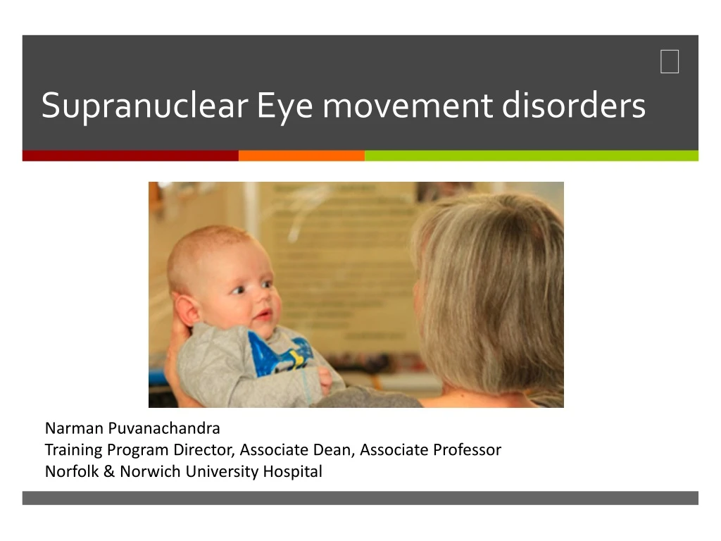
Supranuclear Eye movement disorders. Narman Puvanachandra Training Program Director, Associate Dean, Associate Professor Norfolk & Norwich University Hospital. Learning Objectives. Neuroanatomical basis for Supranuclear / Internuclear control

E N D
Supranuclear Eye movement disorders Narman Puvanachandra Training Program Director, Associate Dean, Associate Professor Norfolk & Norwich University Hospital
Learning Objectives • Neuroanatomical basis for Supranuclear / Internuclear control • Understand the 6 basic types of eye movements • Pathway for each eye movement • How to test each movement • Example of diseases affecting each eye movement • Try not to get confused
Hierarchy of Oculomotor Control Cortical (FEF,PPC,PEF,MT,MST) Superior Colliculus Vestibular Cerebellum Pontine Nuclei Basal Ganglia Supranuclear Affect both eyes together and similarly Brainstem CN Nuclei 3rd/4th Midbrain 6th Pons Medial Longitudinal Fasciculus Nuclear / Inter Affect eyes individually or asymmetrically 3rd, 4th & 6th Cranial nerves Extraocular Muscles Infranuclear
Supranuclear Control Centres of Eye Movement Posterior Parietal (PPC) Parietal Eye Field (PEF) Frontal Eye Field (FEF) Middle (Superior) Temporal (MT / MST) Occipital Lobe Basal Ganglia (BG) Vertical Gaze Centre (Midbrain) riMLF (Rostral Interstitial N of MLF) Superior Colliculus (SC) Interstitial N of Cajal Cerebellum Horizontal Gaze Centre (Pons) PPRF (Paramedian Pontine Reticular Formation) NPH (Nucleus prepositus hypoglossi) Vestibular Nuclei
riMLF riMLF IC IC MIDBRAIN MLF MLF P P R F P P R F PONS
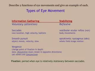
EYE MOVEMENT JEOPARDY WHAT IS ……… ? Compensation for brief head movements = Maintain eye position during sustained head = movements / visual scene moving Hold target on fovea if target slow moving = Place visual target on fovea = Maintain eccentric position of gaze on target = Binocular coordination of foveation = (stereopsis) Vestibular Ocular reflex (VOR) Optokinetic reflex (OKR) Smooth Pursuit (SP) Saccades Gaze holding Vergence
History • Antenatal drugs / infection / medication / drug history • Development history (systemic disorders) • Family History (eyes / neuro / systemic e.g. SCA) • Visual behaviour (do they see, F/F, nyctalopia, photophobia) • Oscillopsia = acquired (if only on head movements = vestibular) • NeuroHx (speech, swallowing, coordination, hearing/tinnitus)
Vestibular Ocular Reflex (VOR) - Pathway Maintain gaze on brief head movement -> Semicircular canals (Inner ear) -> Vestibular Ganglia (Scarpa’s) Internal Auditory Meatus -> Vestibulocochlear Nerve (VIII) -> Vestibular Nuclei (Pons / Medulla) -> Oculomotility Nerves 3,4,6
VOR Supranuclear Control Centres of Eye Movement Cerebellum Vestibular Nuclei
Face turn to the right Vestibular Ocular Reflex (VOR) COMPENSATION FOR BRIEF HEAD MOVEMENTS SLOW EYE MOVEMENT NORMAL = present at birth but some under 6 weeks can lock up in R/L gaze e.g. Face turn to the right ---------------------------- L Ear R Ear VN VN
Vestibular Ocular Reflex (VOR) - Test Doll’s head manoeuvre Rapid but gentle Horizontal / vertical Don’t perform if cervical problems / downbeat nystagmus Normal = Eyes should stay fixed R L
Vestibular Ocular Reflex (VOR) - Test Spinning Baby test Arm’s length / support neck Rotate to right 2-3 revolutions Induces nystagmus fast phase to left Abrupt stop Induces nystagmus fast phase to right Repeat the other way Driven by both VOR & OKR R L
Vestibular Ocular Reflex (VOR) INTERPRETATION Under 45 weeks (normal) -can be normal to lock up on spinning Vestibular lesion = Impaired VOR + catch up saccade on Doll’s head Poor vision / Cerebellar – post-rotational nystagmus > 2-3 beats Systemic syndromes can have poor VOR e.g. CHARGE, Ushers, SCA If patient has a gaze palsy but Doll’s head is preserved then implies a higher level supranuclear disorder
VOR – Clinical Case Acute Peripheral Vestibular insult e.g. Labrynthitis • VOR impaired on ipsilateral head turn; catch up saccade • Unilateral nystagmus fast phase to opposite side (horiz / torsional) • Nystagmus worse on contralateral gaze (Alexander’s Law) • Ipsilateral Head tilt • Tinnitus, Acute painful Vertigo, Hearing loss • No Skew (brainstem / cerebellar) • No other Brainstem / cerebellar signs
Optokinetic Reflex (OKR) Maintain eye position if sustained head move / scene moves Slow Pursuit Component tracks stimulus = Pursuit Pathway Fast Saccadic Component re-fixates = Reflex Saccadic Pathway Normal development Binocular OKR present at birth Monocular OKR present temporal -> nasal at birth But Absent nasal -> temporal At 3 months present both directions monocularly
Optokinetic Reflex (OKR) - test • Optokinetic drum / tape • 50cm from face • Slow rotation • Horizontal both directions • Vertical up and down • Binocularly and monocularly • Normal = • Slow movement in direction of rotation • Fast movement in opposite direction to re-fixate
Optokinetic Reflex (OKR) INTERPRETATION Lack of any OKR = very poor vision (ocular / CVI / DVM) OKR superimposed on child with nystagmus = reassures some vision Fast phase OKR absent = poor saccadic initiation Asymmetry of horizontal OKR binocularly = cortical / pontine lesion (abnormal OKR towards side of cortical / pontine lesion) Vertical binocular OKR worse than horizontal = midbrain disease
Smooth Pursuit - Neuroanatomy Hold target on fovea if slowly moving (<30 degrees / second) Slow Eye Movement 1. IpsilateralParieto-occipital eye field 2. Pontine nuclei 3. Cerebellum (floccular) 4. Vestibular nuclei (contra) 5. Oculomotor nuclei (6,3,4)
SMOOTH PURSUIT Supranuclear Control Centres of Eye Movement Ipsilateral Posterior Parietal (PPC) Parietal Eye Field (PEF) Ipsilateral Middle (Superior) Temporal (MT / MST) Occipital Lobe Cerebellum Vestibular Nuclei Contralateral
Smooth Pursuit – Test Normal Development = jerky (saccadic) smooth pursuit at birth; 3-6 months becomes smoother to larger slow moving targets; gradually smoothsout Test • Horizontally and Vertically with large slow moving toy / mirror • Can use OKR drum / tape (slow component of OKR)
Smooth Pursuit – Clinical Case Cerebellar Flocculus Damage – Alcohol, Drugs, SCA Where you learn to coordinate and remember how to coordinate eye movements “vestibulocerebellum” • Jerky “Saccadic” pursuit all directions • (If only in 1 direction then could be ipsilateralflocculus or ipsilateralParieto-occiptal cortex lesion) • Also effect gaze holding => Gaze evoked nystagmus • Also effect VOR (can’t “remember” coordination) => impaired VOR • Ataxia
Saccades Place visual target on fovea – rapid eye movement Normal Development At birth –hypometric and small secondary saccades after large ones 3 months – less hypometric 1 year – secondary saccades gone
Saccades - Neuroanatomy FEF (contralateral) = Voluntary PEF (contralateral) = Reflexive Cerebellum controls metrics of both Inhibitory burst neurons inhibit contralateral gaze Auditory Somatic Superior Colliculus Retina Visual Cortex Ipsilateral riMLF Ipsilateral PPRF
SACCADES Supranuclear Control Centres of Eye Movement Contralateral Posterior Parietal (PPC) Parietal Eye Field (PEF) Contralateral Frontal Eye Field (FEF) REFLEXIVE VOLUNTARY Ipsilateral SENSORY INTEGRATOR Vertical Gaze Centre (Midbrain) riMLF (Rostral Interstitial N of MLF) Superior Colliculus (SC) Cerebellum Ipsilateral Horizontal Gaze Centre (Pons) CONTROLS METRIC PPRF (Paramedian Pontine Reticular Formation)
Frontal Eye Fields (FEF) Voluntary Saccades Saccade away from target (remember/scan) = Antisaccade Acute deviation of eyes to right and inability to saccade to Left Recovers due to alternative pathways through Superior Colliculus
Saccades - Test Classic Saccadic testing (finger / fist or use toys) • Test Horizontal separately from centre to left / centre to right • Test Vertical top to bottom and back to top • OKN drum includes saccades
Saccades – Clinical Cases • Saccadic Initiation failure – lag of saccade / head thrust = Oculomotor Apraxia (FEF / PEF / Cerebellar Vermis) • Isolated horizontal saccade dysfunction (hypometric / lag) = Pontine lesion (bleed / glioma / Gauchers type 3) • Isolated veritical saccade dysfunction = Midbrain lesion (bleed / Niemann-Pick type C) if Convergence retraction nystagmus / lid retraction / skew = Dorsal Midbrain (Parinauds- aqueduct stenosis / pinealoma) if setting sun sign (tonic downward deviation = raised ICP) • Hypermetric Saccades = Cerebellar lesions
Gaze holding - Neuroanatomy Tonic discharge to maintain eccentric gaze • Horizontal neural integrator - Nucleus prepositushypoglossi (Pons) - Vestibular nuclei (Pons / Medulla) - Vestibulo-cerebellum (Cerebellum) • Vertical neural integrator - Interstitial Nucleus of Cajal (Midbrain) - Flocculus (Cerebellum)
Gaze Holding - Test Observe in primary position first Observe eccentric gaze holding to a toy / target in different positions
Gaze holding - Abnormalities Gaze evoked Nystagmus (GEN) slow drift towards midline and fast corrective saccade Rebound Nystagmus nystagmus beats seen in opposite direction when returning from eccentric gaze to primary position. Often chronic cerebellar disease. Causes – anti-convulsants / drugs - Cerebellar disease (especially if GEN in all directions) - Pontine lesions (purely horizontal GEN) - Midbrain lesions (purely vertical GEN)
Vergence - Neuroanatomy Poorly understood Midbrain / Superior Colliculus Convergence and Divergence neurons Normal Development - start with slight divergence - 2-4 months convergence establishes - Initially driven by accommodation then retinal disparity
Vergence - Test Large visual target brought towards nose 20 PD Base out test from around 6 months
CONVergence - Abnormalities Convergence Excess – Near > Distance Esotropia; bifocals / surgery Convergence Spasm – typically non-organic Convergence insufficiency – common, benign, use exercises / prism Convergence paralysis – rare; rarely isolated, Scan (midbrain)
DIVergence- Abnormalities Divergence Excess –Exotropia Distance > Near; either true or simulated (accommodation / fusion) Divergence insufficiency –v.rare distance esotropia, if other signs scan Divergence paralysis –v.rare. Scan (midbrain / brainstem)
PPRF lesion = Ipsilateral Saccadic Gaze Palsy Ipsilateral Saccadic failure Contralateral gaze evoked nystagmus Acute lesion contralateral gaze deviation riMLF riMLF IC IC MLF MLF P P R F P P R F
MLF lesion = InternuclearOphthalmoplegia On Saccade to contralateral side: Ipsilateral Adduction deficit Contralateral Abduction nystagmus riMLF riMLF IC IC MLF MLF P P R F P P R F
Bilateral MLF lesion Bilateral InternuclearOphthalmoplegia Exotropia Bilateral Adduction deficit Bilateral Abduction nystagmus riMLF riMLF IC IC MLF MLF P P R F P P R F
PPRF & MLF One & a Half Syndrome Ipsilateral Saccadic Gaze Failure On contralateral gaze Failure of adduction ipsilateral eye Preserved Abduction contralateral eye riMLF riMLF IC IC MLF MLF P P R F P P R F
Large Pontine lesion Bilateral Horizontal Gaze Palsy Can be associated with Progressive Scoliosis (ROBO3 gene mutation) riMLF riMLF IC IC MLF MLF P P R F P P R F
Dorsal MIDBRAIN LESION UpGaze Palsy (pursuit / saccade) (overcome by VOR unless severe) Skew Deviation Lid Retraction Convergence Retraction Nystagmus Light / Near Dissociation +/- loss of Convergence riMLF riMLF IC IC MLF MLF P P R F P P R F
Progressive Supranuclear Palsy • Progressive Neuro-degenerative disease (tauopathy) affecting • Basal Ganglia • Midbrain (pons spared) • Cortex • Cerebellum • + rigidity, ataxia, pseudobulbar palsy etc • Resembles Parkinson’s but no tremor • Classically develop a Supranuclear Palsy with • Impaired voluntary vertical saccades • Downgaze first affected • Square-wave jerks • Poor convergence
Supranuclear Control Centres of Eye Movement Posterior Parietal (PPC) Parietal Eye Field (PEF) Frontal Eye Field (FEF) Middle (Superior) Temporal (MT / MST) Occipital Lobe BASAL GANGLIA (BG) Superior Colliculus (SC)
Basal Ganglia PPC FEF Parkinson’s Disease Progressive loss of dopaminergic cells in the substantianigra Impaired Voluntary Saccades Then Reflexive saccades + then Slow pursuit too Also reduced vergence MT / MST Basal Ganglia CN SN SC Caudate Nucleus Substantia Nigra
Neuroanatomical centres REFLEXIVE CONTRALATERAL SACCADES & IPSILATERAL PURSUIT VOLUNTARY CONTRALATERAL SACCADES Posterior Parietal (PPC) Parietal Eye Field (PEF) Frontal Eye Field (FEF) PAUSE ON SUPERIOR COLLICULUS Middle (Superior) Temporal (MT / MST) Occipital Lobe Basal Ganglia (BG) IPSILATERAL PURSUIT Ipsilateral Vertical Gaze Centre (Midbrain) Superior Colliculus (SC) riMLF (Rostral Interstitial N of MLF) SENSORY INTEGRATOR Interstitial N of Cajal Cerebellum Ipsilateral Horizontal Gaze Centre (Pons) CONTROLS METRIC SMOOTH / SACCADE / VOR / VERGENCE PPRF (Paramedian Pontine Reticular Formation) NPH (Nucleus prepositus hypoglossi) Vestibular Nuclei VOR control & Pursuit path
6 Types of Eye Movement • Vestibular Ocular Reflex (VOR) • For brief head turns • Vestibular stimulation leads to Contralateral Gaze • Doll’s Head / Spinning baby • e.g. acute labrynthitis • Optokinetic Reflex (OKR) • Sustained head turn / moving visual scene • Slow phase pursuit • Fast re-fixation saccade • OKN Drum • e.g. good test for vision • Smooth Pursuit (SP) • Hold slow target on fovea • IpsiltateralParieto-occipital • Via Cerebellum / Vestibular • Slow target / OKN drum • e.g. Cerebellar Flocculitis • Saccadic • Place visual target on fovea • Contralateral FEF (volunt) / PEF (reflexive) • Via S.Colliculus / Basal G • Ipsilateral PPRF / riMLF • Saccadic testing / OKN • e.g. Frontal CVA, PPRF lesion • Gaze Holding • Hold target eccentric gaze • NPH (horizontal) • INC (vertical) • + Cerebellum • Hold eccentric gaze • e.g. Gaze Evoked Nystagmus • Vergence • Binocular control (stereo) • Midbrain • Superior Colliculus (SC) • Convergence / Divergence • e.g. DI / DP / DE / CI / CP / CE