Download
1 / 64
640 likes | 847 Views
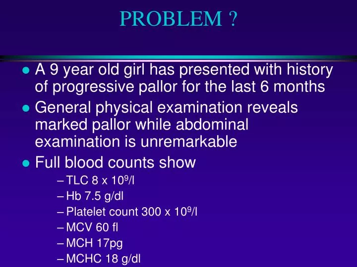
PROBLEM ?. A 9 year old girl has presented with history of progressive pallor for the last 6 months General physical examination reveals marked pallor while abdominal examination is unremarkable Full blood counts show TLC 8 x 10 9 /l Hb 7.5 g/dl Platelet count 300 x 10 9 /l MCV 60 fl

E N D
PROBLEM ? • A 9 year old girl has presented with history of progressive pallor for the last 6 months • General physical examination reveals marked pallor while abdominal examination is unremarkable • Full blood counts show • TLC 8 x 10⁹/l • Hb 7.5 g/dl • Platelet count 300 x 109/l • MCV 60 fl • MCH 17pg • MCHC 18 g/dl
PROBLEM ? • What is the most likely diagnosis ? • What investigations you will carry out to confirm the diagnosis ? • Out line management.
IRON DEFICIENCY ANAEMIA Iron deficiency anaemia is the most common of all the anaemias encountered in clinical practice. Yet it is most often mismanaged.
HYPOCHROMIC MICROCYTIC ANAEMIA • Iron deficiency anaemia • Thalassaemia trait • Anaemia of chronic disorder • Sideroblastic anaemia
IRON BALANCE Iron is the most abundant metal in human body, about 3.5 gm in an adult man; yet the body rigorously conserves it like a trace element.
IRON CYCLE Hb synthesis & Erythropoiesis Intestinal absorption 1 mg/day Plasma iron 13-32 umol/l RBC 2.3 gm Loss 1 mg/day Stores 1 gm RBC destruction & Hb catabolism
TOTAL BODY IRON • Adult male (50 mg/kg) 3.5 gm • Adult female (35 mg/kg) 2.5 gm • Hb 2.3 gm • Stores 1.0 gm • Mb 0.14 gm • Enzymes 0.06 6m
IRON BALANCE • Daily loss 1-2 mg • Average daily in-take 10-15 mg • Normally absorbed (10%) 1-2 mg • Enhancement in deficiency 3-5 mg (20-25% 0f in-take)
IRON ABSORPTION - 1 Cells regulate iron acquisition through post- transcriptional control of apoferritin and transferrin receptor synthesis. mRNA of both proteins contain iron responsive elements (IRE) capable of binding iron regulatory proteins (IRP) 1 & 2. Binding of these proteins has opposing effects on two mRNAs.
IRON ABSORPTION - 2 Transferrin receptor synthesis is directly influenced by the rate of erythropoiesis and indirectly by amount of storage iron (ferritin)
IRON ABSORPTION - 3 • Rate of erythropoiesis/amount of ferritin • Transferrin receptor synthesis • Transferrin synthesis/secretion in bile • Apoferritin and transferrin/mobilferrin in intestinal cell
IRON DEFICIENCY For an individual to become iron deficient, a prolonged period (approximately 6 years), of negative iron balance is required.
IRON DEFICIENCY - STAGES A. Pre - latent iron deficiency Reduction in iron stores without reduction in plasma iron. Serum ferritin & bone marrow iron are reduced. B. Latent iron deficiency Exhaustion of iron stores without reduction in Hb concentration. Plasma iron decreases, TIBC increases and transferrin saturation decreases. C. Iron deficiency anaemia Hb concentration starts declining. Early stage is discovered by chance. Late stage (Hb 8.0 gm) is symptomatic.
IRON DEFICIENCY ANEMIA PATHOGENESIS - 1 Continued negative iron balance Depletion of iron stores Reduction in plasma iron Reduction in Hb synthesis (Increase in free erythrocyte protoporphyrin, hypochromia, microcytosis) Anaemia
IRON DEFICIENCY ANAEMIAPATHOGENESIS - 2 • Negative iron balance results from: • Increases requirements (females) or slow • and steady loss (occult blood loss) • Decreased in-take (poverty, habits) • Combination of the two (most common) • exceeding the physiological limits of absorption • adjustment
IRON DEFICIENCY ANAEMIAPATHOGENESIS - 3 Takes about eight years to develop iron deficiency It takes another 2-3 years to become symptomatic Patients with rapidly developing anaemia seldom become iron deficient as iron is replaced by way of red cell transfusions administered to treat it.
DIAGNOSTIC METHODS - 1 A. Assessment of Iron stores a. Serum ferritin b. Bone marrow iron B. Plasma Iron studies a. Plasma iron b. Serum transferrin (TIBC) c. Transferrin saturation
DIAGNOSTIC METHODS - 2 C. Serum Transferrin receptors D. Red Cell Parameters Early stage a. Red cell free protoporphyrin b. Red cell indices Late stage a. Definite anaemia b. More marked changes in red cell indices and morphology
MANAGEMENT The most important component of effective management for IDA is to find out the cause of chronic negative iron balance and to treat it. Replacement therapy alone will not be able to induce sustained remission.
CAUSES OF IRON DEFICIENCY-1 Increased requirements Decreased in-take Impaired absorption Increased loss (blood loss, 1 ml = 0.5 mg iron )
CAUSES OF IRON DEFICIENCY-3 INFANCY AND CHILD HOOD Prematurity (reduced transfer ) Low birth weight (reduced iron store) Inadequate in-take Increased requirement (with growth) Uncommon vascular anomalies Milk allergy
CAUSES OF IRON DEFICIENCY-4 REPRODUCTIVE FEMALES Menstural disturbances Frequent pregnancies Dietary habits / Pica Hiatus hernia
CAUSES OF IRON DEFICIENCY-5 Hook worms (AD 0.2 ml, NA 0.05 ml / worm / day) Schistosomiasis Ulcerative lesions of GIT Chronic Aspirin ingestion (1-4 ml / day with 02 Tab) Haemorrhoids Neoplasms Runners anaemia (50% of joggers and runners) Nosocomial (ITC 42 ml / day)
INVESTIGATIONS TO DETERMINE THE CAUSE Careful history Thorough physical examination Urine for Hb, haemosidrin, ova Faeces for ova, parasites, occult blood Radiological, Endoscopic examinations Others
CAUSES OF IRON DEFICIENCY-2 Age Sex Socio-economic factors Occupation
REPLACEMENT THERAPY-1 Oral administration is best approach Addition of other elements has no advantage Enteric coating and sustained release reduce absorption Modification of dietary habits greatly improve absorption
RESPONSE Optimal response with 200 mg elemental iron / day For children 1.5-2 mg / kg / day elemental iron Peak reticulocyte (5-10%) between 5th - 10th day Hb at 03 weeks 60% to normal, normal in 2 months Indices normal in 6 months.
INDICATIONS FOR PARENTAL THERAPY Anatomical lesions of upper GIT Functional lesions of upper GIT Rapid loss Extreme intolerance Consistent non-compliance Haemodialysis
CALCULATION OF REQUIREMENT Requirement (mg ) = ( 15 - pt Hb g / dl )x BW (kg)x 3 Either 2 mg I/M daily Or Total dose I/V
REPLACEMENT THERAPY-2 “ Gain in patient acceptance is more important than the reduced absorption of iron “
CAUSES OF FAILURE Incorrect diagnosis Complicating illness Inadequate prescription Continuing loss / malabsorption Non compliance
PREVENTION Premature infants : 02 mg / kg / day at 02 months Infants : 01 mg / kg / day at 04 months Pregnancy : 60 mg ( one Tab of 300 mg ) daily Others : According to loss
IRON DEFICIENCY ANAEMIA Maj Gen Muhammad Ayyub MBBS (Pesh), Ph.D (London), FRC Path (UK), Consultant Haematologist & Commandant Army Medical College, Rawalpindi
β THALASSAEMIA TRAIT • Heterozygous state of β thalassaemia • Usually asymptomatic • Significance of diagnosis • Prenatal counselling • Prenatal diagnosis • Un necessary iron replacement therapy?
LABORATORY INVESTIGATIONS • Blood complete picture • Haemoglobin • Mild anaemia as compared to iron deficiency • Red cell indices • Hypochromic microcytic • Platelet count • Normal • RDW • Normal
LABORATORY INVESTIGATIONS • RBC morphology • Hypochromic microcytic blood picture with mild poikilocytosis and target cells • Definite diagnosis • Haemoglobin electrophoresis • Hb A2 > 3.5%
SIDEROBLASTIC ANAEMIA • Refractory anaemia due to defect in haem synthesis • Defined by presence of > 15% ring sideroblasts in bone marrow out of marrow erythroblasts • Ring sideroblast?
CLASSIFICATION • Hereditary • X linked • Mitochondrial • Autosomal • Acquired • Primary • Myelodysplasia • Secondary • Alcohol, lead, Anti TB, megaloblastic anaemia etc
MANAGEMENT • Blood transfusion • Pyridoxine • Thiamine • Folic acid
DIFFERENTIAL DIAGNOSIS - 1 IDA THAL TR CHR DIS SIDERO Hb. (gm/dl) 8.0 12.0 10.0 6.0 MCV (fl) 74 68 86 77 MCHC (gm/dl) 28 31 32 25 Aniso/Poikilo 1-3+ + + 1-3+ Basophilic stippling 0 2+ 0 2+ Target cells + 5% + 2+ Dimorphism + 0 + 3+
DIFFERENTIAL DIAGNOSIS - 2 IDA THAL TR CHR DIS SIDERO Serum iron N N Transferrin N Saturation N Ferritin N Transferrin N N N receptors
A 42 years old female presented with h/o pallor and generalized weakness and numbness lower limbs for one year. General physical exam revealed marked pallor, red beffy tongue. Abdominal exam is unremarkable. FBC TLC 3.0 x 10⁹/l HB 6.5 g/dl Platelet 100 x 109/l MCV 112 fl MCH 30 pg
INTRODUCTION • Megaloblastic anaemias are a group of disorders characterised by the presence of distinctive morphological appearance (megaloblastic) of erythroid cells in the bone marrow. • Majority of the cases have vitamin B12 or folate dificiency
CAUSES • Vitamin B12 deficiency • Folale deficiency • Defective Vitamin B12 or folate metabolism • Transcobalamin II deficiency • Antifolate drugs • Defects of DNA synthesis • Congenital orotic aciduria • Acquired alcohol, hydroxyurea
Megaloblastic Vitamin B12 deficiency Folate deficiency Non megaloblastic Physiological Pregnancy Infants Pathological Alcohol Liver disease Myeloma MDS Myxodema Reticulocytosis MACROCYTOSIS
MACROCYTOSIS, A PRACTICAL APPROACH • Check history for alcohol and liver disease • Check complete blood counts for evidence of marrow disease • Check B12 and folate levels • Check LFTs and S TSH • Check reticulocyte count
PATHOPHYSIOLOGY Methyl tetrahydrofolate homocysteine B12 Methionine Tetrahydrofolate DHF polyglutamate Tetrahydrofolate polyglutamate 5,10 methylene THF polyglutamate DNA
CLINICAL FEATURES • Anaemia • Jaundice (lemon yellow tint) • Glossitis, angular stomatitis • Peripheral neuropathy • Cardiovascula effects • Features due to thrombocytopenia