Download
1 / 36
360 likes | 787 Views
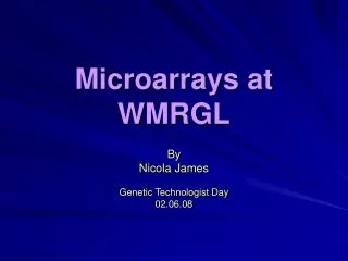
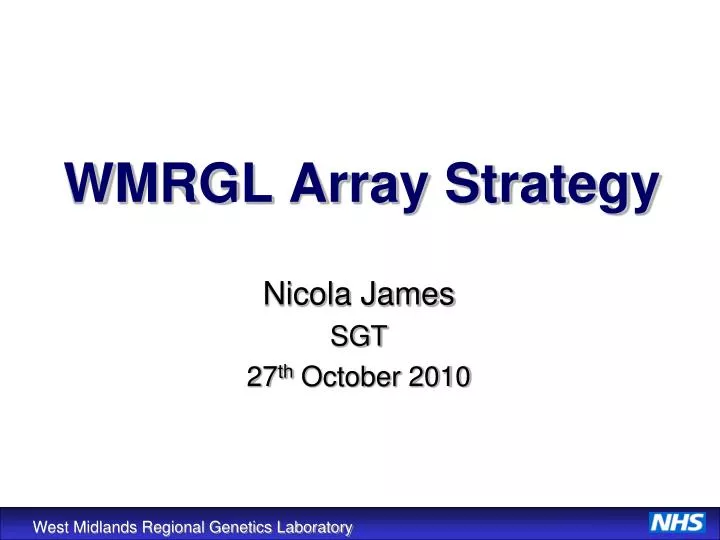
WMRGL Array Strategy. Nicola James SGT 27 th October 2010. Theory re-cap: Array CGH. Ratio. Test Genomic DNA. Reference Genomic DNA. Cot-1 DNA. Genomic clones or oligos spotted on glass slide. DNA gain. DNA loss. Position on Sequence. Referrals for Array CGH.

E N D
WMRGL Array Strategy Nicola James SGT 27th October 2010
Theory re-cap: Array CGH Ratio Test Genomic DNA Reference Genomic DNA Cot-1 DNA Genomic clones or oligos spotted on glass slide DNA gain DNA loss Position on Sequence
Referrals for Array CGH Constitutional: Via Clinical Genetics on a targeted basis until the service is developed, expanded and funded for application to all patients. Prenatal: Pilot PND service to patients with abnormal ultrasound scans including cardiac defects referred via FMT at BWH or selected Fetal Pathology cases. Oncology: Selective leukaemia/solid tumour samples for additional delineation of abnormal karyotypes.
Array Platforms CytoChip Arrays: Diseased focused arrays with extra tiling over regions associated with genetic disorders. New 24 Sure Technology Rapid, reliable aneuploidy screening within 12 hours using picograms of DNA Validated for use with single cells (polar bodies, blastomere biopsies, IVF applications and stem cell research) V3 BAC array 0.5Mb backbone 250Kb subtel. 110 known disorders @ 100Kb Agilent ISCA Various formats 4x44K 8x60K 4x180K 2x10⁵K BAC array 1Mb backbone 100Kb over 97 disease regions BAC array Tiling over regions associated with haematological malignancy
Array Platforms OGT: 2x10⁵K oligo X chromosome array - XLMR identified patients - Agilent printed oligonucleotide array - 97,248 X chromosome probes with exonic bias (1 probe/1.8Kb in exons and 1 probe/15Kb in introns). - Cases analysed using OGT CytoSure Software Affymetrix: 2.7M WG array -Research staff and project work (non-diagnostic) - New whole genome 2.7M array being trialled - SNP array, can detect LOH/acquired isodisomy Future – Gene Titan 96 well plate format arrays!
WMRGL Array CGH Referrals: Protocol Summary Report Blood cultured and DNA extracted FISH/molecular validation studies DNA quality assessment Batch run assignment Result interpretation and analysis Random prime labelling of DNA Image acquisition via laser scanning Labelled DNA purification, assessment and precipitation Slide washing/drying Pre-hybridisation with Cot-1 Overnight hybridisation to array
DNA Quality Control Crude band assessment on gel (select samples) High molecular weight DNA required Nanodrop assessment of optical density: accuracy important for subsequent assays Failed samples: attempt Na+/ethanol precipitation
DNA Labelling DNA labelled with Cy3 and Cy5 (reference DNA also) Reactive water soluble fluorescent dyes with side groups so that they can chemically link to nucleic acids. Cy3 excited at 550nm emits at 570nm (red part of spectrum) Cy5 excited at 649nm emits at 670nm (far red part of spectrum) Scanner can easily distinguish between the two Labelling method specific to platform but theoretically very similar…
Random Prime Labelling Reaction Reaction mix contains high molecular weight genomic DNA, random primers, Cy-dCTPs (BAC)/dUTPs (oligo), dNTPs (lower concentration C/T’s) Catalysed by Klenow enzyme Genomic DNA Extension by Klenow 2 hour or overnight incubation @ 37oC Primer Annealing Heat Denaturation Snap Chill Heat denaturation/addition of EDTA to stop the reaction Labelled DNA needs quantification and purification
Purification + Dye Incorporation Spin 2000g 1min Labelled DNA Nanodrop QC: Concentration >100ng/µl Dye incorporation >3pmol/µl Measure dye incorporation on Nanodrop
Sample Pre-hybridisation Test Cy3 + Reference Cy5 (dye swap: Test Cy5 + Reference Cy3) + + Precipitation with sodium acetate and ethanol Re-suspension Oligo Arrays DNA hydration solution Blocking agent/buffer 95°C denaturation BAC Arrays Hybridisation solution 75°C denaturation
BAC Arrays 1 hour at 37°C Hyb solution made manually Cot 1 DNA: blocks repetitive sequences HS DNA: minimises non-specific binding Formamide: lowers denaturation temp to 75°C Dextran sulphate: bulking agent/volume exclusion % content optimised depending on auto/manual protocol Oligo Arrays 30 mins at 37°C Hyb cocktail contents are “kit based”; constituent reagents not specified unless hazardous. Cot 1 DNA: blocks repetitive sequences 10xblocking agent: minimises non-specific binding ? contains HS DNA 2xhyb buffer: ? bulking agent like DS stabilises labelled DNA during 95°C denaturation Contains oxirane, methyl-, polymer with oxirane, mono[3-[1.3.3.3 - tetramethyl-1 [(-trimethylsiltl) oxy]disiloxanyl]propyl] ether Pre-Hybridisation
CytoChips clamped in place in Tecan station Samples injected into appropriate chamber Automated hybridisation for 21 hours Automated slide washing to remove unbound and non-specifically bound DNA ~ 1 hour 2xSSC, 0.01% SDS 36.5°C 0.1xSSC, 54°C – most stringent 2xSSC, 0.01% SDS 23°C Distilled H20, 23°C Slide drying with compressed nitrogen Manual BAC array method under investigation following equipment problems and reduction in usage BAC Array Hybridisation Tecan HS 4800 Pro Hybridisation Station
Manual array loading onto gasket slide (2x, 4x and 8x formats) Gasket/array slide assembly clamped Hybridised in oven at 65°C for 24hours H G G H F E E F D C C D B A A B GASKET SLIDE ARRAY SLIDE Oligo Array Hybridisation Mirrored!
Manual washing step Kit based wash buffers Stringency optimised for oligo probe chemistry Gasket assembly disassembled in wash 1 Wash 1 at room temperature, 5 mins Wash 2 at 37°C, 1 minute > Hybex incubation system; temp crucial to successful washing > Drying/stabilisation properties of this wash; slides dry when removed Oligo Array Washing
Scanning CytoChips protected in slide holder which slot into scanner Laser scanner simultaneously scans red and green signals Rapid; time depends on platform and scan resolution BAC arrays Scan top and bottom sub arrays separately at 10µm resolution; approx 4 minutes per half prenatal BAC array Oligo arrays Scan whole slide region at 3µm; extract sub array images later in array processing software (feature extraction software for X arrays). Agilent scanner
From this…. raw BAC array image ….to this whole genome view: BAC array …in several complicated mathematical steps
raw oligo array image +ve and –ve controls whole genome view: oligo array
BlueFuse Multi (BlueGnome, Cambridge) Fully automated, based on advanced statistical modelling Software used as a database to store QC information, labelling info, as well as the “experiment” itself. Raw data files inputted into software under patient “experiment” Cases processed using: > specific GAL file (depending on array type and batch) > database annotation file (genomic info that array based on) Array Processing Software
Grid alignment - finding spot centres/array edges/orientation Quantification - calculating raw spot intensities+log2ratios Normalisation - correcting bias in the log2ratio (GC shift, spatial) Exclusion (BAC arrays) - throwing away any bad spots Fusion (BAC arrays) - averaging good replicates Combining (BAC arrays) - combining top + bottom arrays Smoothing – >BAC arrays: Focused platforms can be smoothed in backbone regions differentially to disease specific regions to reduce number of calls that may be difficult to interpret >Oligo arrays: Reduces noise in the array profile so that oligo changes without at least one adjacent supporting oligo are removed (single oligo insufficiently robust to provide reliable information source). Copy number calling – determining regions of copy number change Array Processing Software
BAC Array Copy Number Calling • A significant copy number change must; • Exceed 3 SD from the autosome • Exceed a set significance threshold (log2ratio >+/-0.3) • A single clone exceeding SD threshold of +/-0.3 is significant • To ensure detection of mosaicism; • A region of 3 consecutive clones exceeding SD threshold and • median of only +/-0.26 will also be called as significant • A region of 10+ consecutive clones exceeding SD threshold and • median of only +/-0.20 will also be called as significant • Individual clones in regions of clear copy number change with a different • relative change will be called; • ie. if 4 clones are called as a duplication but the middle clone is • called as a deletion this clone will be called as a deletion
DNA gain DNA loss BAC Array Whole Genome View Log2ratio 0.3 Log2ratio -0.3
Oligo Array Copy Number Calling An aberration must include sufficient oligos to reliably distinguish it from noise. At least 3 oligos are required for any call. Depending on the number of oligos involved, an aberration is required to be separated from the autosome by both a set number of robust standard deviations and a fixed log2 ratio threshold. The larger the number of oligos involved, the smaller the separation required. A log2 ratio of at least 0.3 is required for smaller aberrations. Therefore, a 50 oligo aberration requires very little separation to be called as significant allowing reliable detection of larger low level mosaic aberrations.
DNA gain DNA loss Oligo Array Whole Genome View
BAC arrays -Grid position -Raw image for washing artefacts -Confidence scores (high A+B) -%BACs included ≥95%. Measure of technical reproducibility -SD autosome ideally between 0.03 and 0.07. Measure of biological reproducibility (noise) Oligo arrays -Grid position -Raw image for washing artefacts -Derivative Log Ratio (DLR); robust noise measurement even in presence of highly aberrant samples (differences in log 2 ratio between adjacent oligos) -SD autosome; higher than BAC arrays (significantly more data points!) Post Processing Batch Array QC Identifies potential issues across a batch which can then be investigated…
Using a range of online analysis tools: >Database of Genomic Variants: summary of structural variation in the human genome (healthy control individuals) >Ensembl: genome view, mapped genes, tilepath clones >DECIPHER: clinical resource with consented case information >CytoChip user group: email correspondence to discuss results >ECARUCA >SUSPECTS, Top Gene etc. Investigate regions called by the software and classify individual calls as appropriate (pathogenic, benign CNV, rare CNV etc). Care! Databases constantly updating! Also need to ensure aligned to same genomic annotation as array based on. Analysis Overview
Cytogenetic Based Validation FISH (fluorescent in situ hybridisation) Frontline validation technique BAC clones ordered from either Roswell Park Cancer Institute (RPCI) and labelled in house by random prime labelling, or pre labelled from BlueGnome. BAC Rack database to catalogue probes and give set up and mapping information. Advantages; Inexpensive Large stock of labelled probe produced Quick Technique already in place (have a FISH department) Provides positional information in proband and parents Disadvantages; Difficult to validate small duplications (<1Mb)
DNA Based Validation Alternative microarray platform Affymetrix Agilent/OGT X tiling path MLPA Commercial kits (Mental Retardation; 1p-deletion, Williams, Smith-Magenis, Miller-Dieker, DiGeorge, Prader-Willi) Home made assays (lots in house in molecular section) Quantitative PCR Would need to work up an individual assay for each case to look for particular imbalance
“oliGO”ing forward! • Higher resolution oligo arrays to detect smaller imbalances are now • platform of choice for the majority of referrals. • Frontline platform now 8x60K which is higher resolution and • significantly cheaper than V3 BAC array due to multi sub array design. • V3 BAC array costs £250 per patient more than the cost per patient of • an 8x60K array. • A single batch run of 8x60K arrays (4 arrays, 32 patients) is • approximately £8,000 cheaper than running 32 V3 BAC arrays • over 4 batch runs!!! In other words, 1 week’s work vs 4 week’s • work……no brainer?!!! • Tecan HS 4800 Pro unreliable as of late – manual BAC array method for • prenatal referrals is a current priority.
Future Development Challenges • Increase in number of referrals (frontline service) • Service re-design to accommodate high throughput • Automation and technical back-up (no failure!) • Staff re-training • IT capacity: analysis and data storage • Interpretation and validation • PND service considerations – trial oligo platform?
Manual Max 16 to 32 patients per week (1 to 2 GTs) Lots of sample tubes! Staggered processing due to equipment capacity Batch processing in “small” batches Automated 96 well plate based Potential for >96 patients per week Reduced “hands on” time Still need for individual sample QC Equipment requirements are considerable! Need service contracts too! Batch processing of many results simultaneously Protocol Overhaul!
Equipment Requirements Multichannel pipettes and liquid handling robot? Facilitate 96 well plate labelling assays 8-channel NanoDrop Spectrophotometer
Equipment Requirements Vacuum centrifuge: filter column plates Thermal cycler Plate centrifuge
Ozone Monitoring • Ozone monitoring ? “Effects of Ozone Exposure during Microarray Posthybridization Washes and Scanning”: Signature Genomics: Journal of Molecular Diagnostics, Vol. 11, No. 6, November 2009 • No internal WMRGL ozone monitoring to date Potentially need ozone scrubbers/cabinets?
Summary Array CGH is a high resolution technique, but manual methods are still time consuming and relatively expensive. Array costs are decreasing with new format platforms that allow multiple hybridisations per slide (e.g. 8x60K oligo) – next step will be to introduce automation (plate based labelling assays). Balanced rearrangements not detected. Validation required to confirm results. Distinguishing between rare polymorphisms and genuine pathogenic imbalances difficult. Higher resolution platforms means more data and therefore more in depth analysis!