Download
1 / 32
320 likes | 330 Views
The Foot As a Functional Unit. Dr. Fadel Naim Orthopedic Surgeon El- Wafa Medical Rehab. And Surgical Hospital. The Foot As A Weight-bearer And A Lever. The foot has two important functions: To support the body weight

E N D
The Foot As a Functional Unit Dr. Fadel Naim Orthopedic Surgeon El- Wafa Medical Rehab. And Surgical Hospital
The Foot As A Weight-bearer And A Lever • The foot has two important functions: • To support the body weight • To serve as a lever to propel the body forward in walking and running. • If the foot possessed a single strong bone, instead of a series of small bones, it could fulfill those functions • Could not adapt itself to uneven surfaces • The forward propulsive action would depend entirely on the activities of the gastrocnemius and soleus muscles. • Because the lever is segmented with multiple joints • The foot is pliable and can adapt itself to uneven surfaces. • The long flexor muscles and the small muscles of the foot can • Exert their action on the bones of the forepart of the foot and toes • Greatly assist the forward propulsive action of the gastrocnemius and soleus muscles
The Arches of the Foot • A segmented structure can hold up weight only if it is built in the form of an arch. • The foot has three such arches, which are present at birth: • Medial longitudinal • Lateral longitudinal • Transverse arches • In the young child, the foot appears to be flat because of the presence of a large amount of subcutaneous fat on the sole of the foot
On examination of the imprint of a wet foot on the floor made with the person in the standing position, one can see that following structures are in contact with the ground • The heel • The lateral margin of the foot • The pad under the metatarsal heads • The pads of the distal phalanges
The medial margin of the foot, from the heel to the first metatarsal head, is arched above the ground because of the important medial longitudinal arch. • The pressure exerted on the ground by the lateral margin of the foot is greatest at the heel and the fifth metatarsal head • Least between these areas because of the presence of the low-lying lateral longitudinal arch.
The transverse arch involves the bases of the five metatarsals and the cuboid and cuneiform bones. • This is, in fact, only half an arch • The foot has been likened to a half-dome • when the medial borders of the two feet are placed together, a complete dome is formed. • The body weight on standing is distributed through a foot via: • The heel behind • Six points of contact with the ground in front • The two sesamoid bones under the head of the first metatarsal • The heads of the remaining four metatarsals
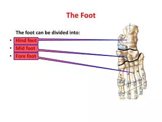
The Bones of the Arches • Medial longitudinal arch: • This consists of: • The calcaneum • The talus • The navicular bone • The three cuneiform bones • The first three metatarsal bones
The Bones of the Arches • Lateral longitudinal arch: • This consists of: • The calcaneum • The cuboid • The fourth and fifth metatarsal bones
Transverse arch: • This consists of: • The bases of the metatarsal bones • The cuboid • The three cuneiform bones
Mechanisms of Arch Support • The shape of the stones: • The most effective way of supporting the arch is to make the stones wedge shaped, with the thin edge of the wedge lying inferiorly. • Keystone • The important stone that occupies the center of the arch
Mechanisms of Arch Support • The inferior edges of the stones are tied together. • Interlocking the stones or binding their lower edges together with metal staples. • Effectively counteracts the tendency of the lower edges of the stones to separate when the arch is weight-bearing.
Mechanisms of Arch Support • The use of the tie beams: • When the span of the bridge is large • The foundations at either end are insecure • A tie beam connecting the ends effectively prevents separation of the pillars and consequent sagging of the arch
Mechanisms of Arch Support A suspension bridge: • Multiple supports suspending the arch from a cable above the level of the bridge
Maintenance of the Medial Longitudinal Arch • Shape of the bones: • The sustentaculum tali holds up the talus • The concave proximal surface of the navicular bone receives the rounded head of the talus • The slight concavity of the proximal surface of the medial cuneiform bone receives the navicular. • The rounded head of the talus is the keystone in the center of the arch
Maintenance of the Medial Longitudinal Arch • The inferior edges of the bones are tied together by the plantar ligaments • Which are larger and stronger than the dorsal ligaments. • The most important ligament is the plantar calcaneonavicular ligament • The tendinous extensions of the insertion of the tibialis posterior muscle play an important role in this respect.
Maintenance of the Medial Longitudinal Arch • Tying the ends of the arch together are • The plantar aponeurosis • The medial part of the flexor digitorum brevis • The medial part of the flexor digitorum longus • The abductor hallucis • The flexor hallucis longus • The flexor hallucis brevis
Maintenance of the Medial Longitudinal Arch • Suspending the arch from above are • The tibialis anterior • The tibialis posterior • The medial ligament of the ankle joint.
Maintenance of the Lateral Longitudinal Arch • Shape of the bones: • Minimal shaping of the distal end of the calcaneum and the proximal end of the cuboid. • The cuboid is the keystone • The inferior edges of the bones are tied together by • The long and short plantar ligaments • The origins of the short muscles from the forepart of the foot
Maintenance of the Lateral Longitudinal Arch • Tying the ends of the arch together are • The plantar aponeurosis • The abductor digiti minimi • The lateral part of the flexor digitorum longus and brevis. • Suspending the arch from above are • The peroneus longus and the brevis
Maintenance of the Transverse Arch • Shape of the bones: • The marked wedge shaping of the cuneiform bones and the bases of the metatarsal bones • The inferior edges of the bones are tied together by • The deep transverse ligaments • The strong plantar ligaments • The origins of the plantar muscles from the forepart of the foot • The dorsal interossei • The transverse head of the adductor hallucis
Maintenance of the Transverse Arch • Tying the ends of the arch together • The peroneus longus tendon. • Suspending the arch from above are • The peroneus longus tendon • The peroneus brevis.
The tibialis anterior • The peroneus longus • The small muscles of the foot • Play no important role in the normal static support of the arches • They are commonly totally inactive. • However, during walking and running all these muscles become active • Standing immobile for long periods, especially if the person is overweight, places excessive strain on the bones and ligaments of the feet and results in fallen arches or flat feet • Athletes, route-marching soldiers, and nurses are able to sustain their arches provided that they receive adequate training to develop their muscle tone.
Pes planus (flat foot) • A condition in which the medial longitudinal arch is depressed or collapsed. • As a result, the forefoot is displaced laterally and everted. • The head of the talus is no longer supported • The body weight forces it downward and medially between the calcaneum and the navicular bone. • When the deformity has existed for some time • The plantar, calcaneonavicular, and medial ligaments of the ankle joint become permanently stretched, and the bones change shape. • The muscles and tendons are also permanently stretched. • The causes of flat foot are both congenital and acquired.
Pes cavus (clawfoot) • a condition in which the medial longitudinal arch is unduly high. • Most cases are caused by muscle imbalance, in many instances resulting from poliomyelitis.
The Propulsive Action of the Foot • Walking • As the body weight is thrown forward, the weight is born successively on the lateral margin of the foot and the heads of the metatarsal bones. • As the heel rises, the toes are extended at the MTP joints • The plantar aponeurosis is pulled on, thus shortening the tie beams and tightening the longitudinal arches. • The "slack“ of the long flexor tendons is taken up, thereby increasing their efficiency. • The body is then thrown forward by • The actions of the gastrocnemius and soleus (and plantaris) on the ankle joint, using the foot as a lever • The toes being strongly flexed by the long and short flexors of the foot, providing the final thrust forward. • The lumbricals and interossei contract and keep the toes extended so that they do not fold under because of the strong action of the flexor digitorum longus. • In this action, the long flexor tendons also assist in plantar flexing the ankle joint.
The Propulsive Action of the Foot • Standing immobile • The body weight is distributed via the heel behind and the heads of the metatarsal bones in front including the two sesamoid bones under the head of the first metatarsal • Running • When a person runs, the weight is borne on the forepart of the foot, and the heel does not touch the ground. • The forward thrust to the body is provided by the mechaisms described for walking
GENU RECURVATUM • Hyperextension of the knee joint, is found in babies who have had a breech presentation with extended legs • No treatment is required, because the legs return to normal within a few weeks.
TALIPES • Talipes (club foot) often is caused by abnormal position or restricted movement of the fetus in utero. • A small number of cases may be caused by muscle paralysis associated with spina bifida. • The different types are named according to the position of the foot. • In talipes equinovarus • The foot is plantar flexed at the ankle joint • Inverted at the midtarsal joints • The conditions may be unilateral or bilateral • They require orthopedic treatment • Talipes calcaneovalgus • A form of club foot in which • The foot is dorsiflexed at the ankle joint • Everted at the midtarsal joints.
METATARSUS VARUS • Metatarsus varus is a common condition in which the forefoot is adducted on the rear part of the foot. • Correction may be accomplished by manipulation followed by splinting
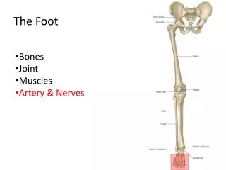
ARTERIAL PALPATION • Every health professional should know the precise position of the main arteries within the lower limb • The femoral artery: • At a point midway between the anterosuperior iliac spine and the symphysis pubis • The popliteal artery • Can be felt by gentle palpation in the depths of the popliteal space provided that the deep fascia is fully relaxed by passively flexing the knee joint • The dorsalis pedis artery • Lies between the tendons of extensor hallucis longus and extensor digitorum longus, • Midway between the medial and lateral malleoli on the front of the ankle • The posterior tibial artery • The pulsations of the artery can be felt midway between the medial malleolus and the heel
ARTERIAL OCCLUSIVE DISEASE OF THE LEGIntermittent Claudication • Common in men. • Ischemia of the muscles produces a cramp-like pain with exercise. • The patient is forced to stop walking after a limited distance because of the intensity of the pain. • With rest, the oxygen depletion is corrected and the pain disappears. • However, on resumption of walking, the pain recurs.