Download
1 / 34
360 likes | 404 Views
Thoracolumbar Fractures. Classification Models Fracture Management Case Illustrations. Kevin Chao, MD Stanford Neurosurgery. Classification Models. Denis Three-column ANATOMIC biomechanical model Accounts for mechanism of injury No rigid guidelines for treatment Magerl/ AO Spine

E N D
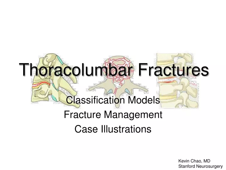
Thoracolumbar Fractures Classification Models Fracture Management Case Illustrations Kevin Chao, MD Stanford Neurosurgery
Classification Models Denis • Three-column ANATOMIC biomechanical model • Accounts for mechanism of injury • No rigid guidelines for treatment Magerl/ AO Spine • MECHANISTIC model • Detailed characterization of fracture subtypes Thoracolumbar Injury Classification and Severity Score (TLICS) • Accounts for 1) fracture morphology, 2) neurologic deficit, and 3) posterior complex status • Point-based system guides intervention (TLICS > 4)
Denis Three Column Model Posterior SSL/ISL Posterior arch Facet capsule Ligamentum flavum Anterior ALL Ant annulus Ant wall VB Middle PLL Post annulus Post wall VB
Denis Series Outcomes(412 pts) !! Denis gives NO RIGID PARAMETERS for treatment
TLICS TOTAL ___
ASIA Scale Poor prognosis Good prognosis
Spine Fracture Approach • Management • Surgery vs no surgery • Goals? • Brace? • Activity restrictions • Follow up • Imaging • Rehab Assessment • Mechanism of injury • Neuro exam • Imaging • Levels • Bone vs soft tissue • Dynamic vs static • Vessels? • Degree of instability
Stable or Unstable? Overall degree of instability 1st degree: Mechanical instability 2nd degree: Neurological instability 3rd degree: Both
Stable or Unstable? STABLE Minimal anterior column wedge Above T8 if ribs and sternum intact Seat-belt type injuries without neurologic deficit UNSTABLE > 50% height loss > 20°angulation > 50% canal compromise* Neurologic deficit Progressive kyphosis
Fracture Management Goals: • Mechanical stabilization • Prevention of secondary neurologic injury • [ Decompression, if needed ] !! Instrumentation only serves as a bridge to fusion (or ligament healing)
35M paragliding accident • Motor • Right hip flexion pain-limited weakness (otherwise full strength) • Normal rectal tone • Sensory • Right thigh to knee completely numb • Left knee and shin partly dumb • Saddle anesthesia • Reflexes • Diminished at knees and ankles • No clonus L2 3rd degree instability TLICS 5 (2+3+0) ASIA D
Two-stage procedure Stage 1 Segmental instrumentation T12-L4 Decompression Posterolateral fusion
Two-stage procedure Stage 2 Lateral corpectomy Interbody cage
Post op result • Motor • Improved hip flexion • Able to walk • Sensory • Unchanged • Bracing • TLSO • Follow up • - 4 week repeat X rays
Teaching points • Recognize cauda equina syndrome • Define surgical goals • Many approach options (P, A/P, L/P) • Lateral approach technique • No abd surgery exposure needed • L3-T12 (below L3 often limited by iliac crest) • Rib resection +/- chest tube may be needed • Lumbar lordotic curve significant load bearing in middle and posterior columns
42M fell from tree • Motor • Full strength • Normal rectal tone • Sensory • Intact to LT, proprioception, pin prick • Reflexes • Normal at knees and ankles • No clonus T12 1st degree instability TLICS 7 (4+0+3) ASIA E
Sag CT recon Facet disruption MR Sag STIR Disc extrusion Ligament disruption MR Axial T2 FS Canal hematoma Facet disruption
Post op result • T11-L2 posterolateral fusion • Motor • Intact • Sensory • Intact • Bracing • TLSO • Follow up • - 6 week repeat X rays pending
Teaching points • Look beyond static image: What was the mechanism of injury? • Ligamentous injury >> bony injury • Ligamentous seat-belt-type fracture management options: • Open surgical instrumentation/fusion • Internal bracing (i.e. percutaneous instrumentation) • Bracing ?
22M motorcycle crash • Motor • 2/5 hip flexion and knee extension • 0/5 below knee • diminished rectal tone • Sensory • Diminished sensation to light touch below knee • Reflexes • None at knees and patella • No clonus 3rd degree instability TLICS 9 (3+3+3) ASIA C
1st attempt at surgery: Aborted due to sacral hemorrhage Wound packed Pelvic binder placed Sacral vessels embolized Transfused pRBC, FFP, plts Returned to OR 2 days later…
Post op result • L1-L5 segmental instrumentation and posterolateral fusion • Correction of fracture-dislocation using Wilson frame and reduction screws • Motor • unchanged • Sensory • some ROF below knees • Bracing • TLSO • Follow up • - 6 week repeat X rays Not yet
Teaching points • Fracture-dislocations lead to majority of neurologic deficits from spine traumas (~50%) • Recognize other trauma injuries • Many spine fractures are URGENT (treat within 48 hours). Very few are EMERGENT (treat < 12 hours). • Wait for hemodynamically stability AMAP • Know fracture pattern/ anatomy preop • Be prepared for other injuries (thecal sac/ nerve roots, vascular, ureters, bowel, etc)
Teaching points • Can reduce some fractures with special OR tables (Wilson frame, Axis tilt, Jackson prone) • Reduction screws can be very helpful Axis-Jackson Table Reduction screw Wilson frame
Final point • TP fractures are not always benign • L4-5 TP fractures associated with lumbosacral plexus injury • T1-2 TP fractures associated with brachial plexus injury
References • Denis F. The Three Column Spine. Spine 1983; Vol. 8, No 8: 817-831 • Classic historic paper with simple classification system • No rigid parameters for treatment • Patel A, Vaccaro A. Thoracolumbar Spine Trauma Classification. J Am Acad Orthop Surg 2010;18: 63-71 • New TLICS classification point system to guide treatment • http://www.aospine.org/ • Pocket cards and protocols