Download
1 / 41
470 likes | 946 Views
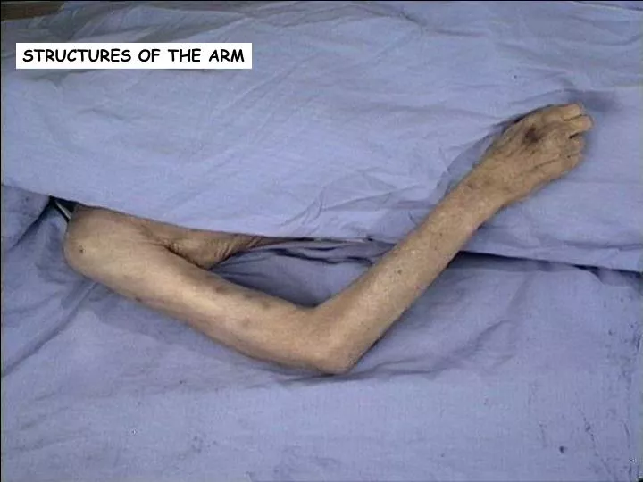
STRUCTURES OF THE ARM. !!!! IMPORTANT !!!!. BEFORE making any incisions – make certain you remember the course of the superficial veins of the upper limb!!!!!!!!!!!!!!!!!!!!!!!! LEAVE THE SUPERFICIAL VEINS INTACT!!!!!!!!!

E N D
!!!! IMPORTANT !!!! BEFORE making any incisions – make certain you remember the course of the superficial veins of the upper limb!!!!!!!!!!!!!!!!!!!!!!!! LEAVE THE SUPERFICIAL VEINSINTACT!!!!!!!!! Remember that these superficial veins are somewhat conspicuous in the living individual and are frequently the sites for drawing blood and injecting medications. BUT, in the cadaver, the superficial veins are empty and generally cannot be seen through the skin. It is my opinion that you are most likely to save these veins if you begin the removal of skin and superficial fascia from the most distal site, i.e., the dorsal hand or carpus. The veins in this region drain into the basilic and cephalic veins. So, once these veins are found in this region, it is much easier to simply follow them proximally as you continue dissection.
Basilic vein Cephalic vein Median cubital vein
Cephalic vein in the arm Cephalic vein in the forearm Basilic vein in the arm Median antebrachial vein Median cubital vein
Cephalic vein emptying into the axillary vein Cephalic vein in the arm Cephalic vein in the forearm
SKIN INCISIONS • Using the next slide for reference, make the following skin incisions with the cadaver in the supine position. • From the jugular notch A along the clavicle and across the acromion B to a point about 10 cm distal to the acromion. • From A to the xiphisternal junction C. • From C laterally to the table. • At about mid-arm, make a complete circular incision. • At the level of the wrist make another circular incision. • Join these two circular incisions with a longitudinal one on the lateral aspect of the upper limb, that extends to the cut that is distal to the acromion.. Reflect the skin of the arm and forearm and remove it completely. DO NOT damage the superficial veins and cutaneous nerves in the superficial fascia.
Skin Incisions B C A B
The cephalic vein is usually best identified first at its most proximal site - piercing the fascia of the deltopectoral triangle. This vein passes along the lateral aspect of the arm and enters this deltopectoral groove or triangle between the pectoralis major and deltoid mm..
Obviously, to get to this site, you must clean the anterior surface of the pectoralis major muscle and define its borders. Recall that the pectoralis major muscle has two heads: a clavicular and sternocostal head. The fibers of the two heads converge and insert into the lateral lip of the bicipital groove of the humerus.
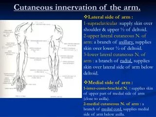
You should be able to identify the various cutaneous nerves that pass through the superficial fascia to supply the skin (but will probably not be able to save many of them as you continue with your dissection.) Medial brachial cutaneous nerve
Reflect the pectoralis major m. laterally (only if you have been specifically told to do so.)
Identify the tendon of the long head of the biceps brachii m..
Musculocutaneous nerve going through the coracobrachialis muscle Branches of the musculocutaneous nerve going to the biceps brachii muscle
The musculocutaneous nerve terminates as the lateral antebrachial nerve which passes lateral to the tendon of the biceps muscle and enters the forearm, providing cutaneous sensation to that region.
Median nerve traveling from the axilla to the cubital fossa. This nerve generally lies medial to the brachial artery throughout its course in the arm. Remember that it does not innervate any muscles in the arm.
Identify the ulnar nerve between the axilla and the posterior aspect of the medial epicondyle of the humerus. Like the median nerve, the ulnar nerve does not supply any muscles in the arm.
Remember that the axillary artery becomes the brachial artery at the inferior border of the teres major muscle.
Identify the profunda brachii artery that arises near the beginning of the brachial artery and runs with the radial nerve to supply the muscles of the posterior compartment of the arm.
You may also see collateral arteries (which provide collateral circulation around the elbow joint) arising from the brachial artery. Here the probe is pointing to the superior ulnar collateral artery. Usually this vessel travels with the ulnar nerve posterior to the medial epicondyle.
Note that between the brachialis and brachioradialis mm., you should find the radial nerve.
Now, to look at the muscles of the posterior arm, it is best to rotate the arm medially and reflect the deltoid m.. BUT do NOT reflect the deltoid m. until you are told to do so.
From this position the medial head of the triceps brachii m. cannot be seen well unless the lateral head has been cut and reflected – which you probably do NOT want to do.
From this position though, you can see the exposed length of the radial nerve. Note how it is direct contact with the humerus (radial or spiral groove). You should be able to see how this nerve can be easily damaged with mid-shaft fractures of the humerus.
You should be able to see some branches of the radial nerve reaching the various heads of the triceps brachii m..
Also note how the profunda brachii artery is traveling with the radial nerve in this region.