Download

1 / 57
690 likes | 1.02k Views
3. Cells and Tissues. Epithelial Tissues. Locations Body coverings Body linings Glandular tissue Functions Protection Absorption Filtration Secretion. Body Tissues. Tissues Groups of cells with similar structure and function Four primary types Epithelial tissue (epithelium)

E N D
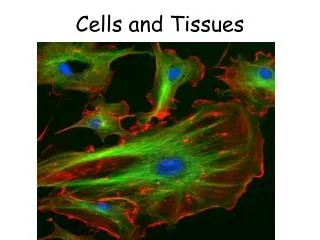
3 Cells and Tissues
Epithelial Tissues Locations Body coverings Body linings Glandular tissue Functions Protection Absorption Filtration Secretion
Body Tissues Tissues Groups of cells with similar structure and function Four primary types Epithelial tissue (epithelium) Connective tissue Muscle tissue Nervous tissue
Epithelium Characteristics Cells fit closely together and often form sheets The apical surface is the free surface of the tissue The lower surface of the epithelium rests on a basement membrane Avascular (no blood supply) Regenerate easily if well nourished
Apical surface Basal surface Simple Apical surface Basal surface Stratified (a) Classification based on number of cell layers Figure 3.17a
Classification of Epithelia Number of cell layers Simple—one layer Stratified—more than one layer
Apical surface Basal surface Simple Apical surface Basal surface Stratified (a) Classification based on number of cell layers Figure 3.17a
Classification of Epithelia Shape of cells Squamous flattened Cuboidal cube-shaped Columnar column-like
Simple Epithelia Simple squamous Single layer of flat cells Location - usually forms membranes Lines body cavities Lines lungs and capillaries Functions in diffusion, filtration, or secretion in membranes
Air sacs of lungs Nucleus of squamous epithelial cell Nuclei of squamous epithelial cells Basement membrane Photomicrograph: Simple squamous epithelium forming part of the alveolar (air sac) walls (185×). (a) Diagram: Simple squamous Figure 3.18a
Simple Epithelia Simple cuboidal Single layer of cube-like cells Locations Common in glands and their ducts Forms walls of kidney tubules Covers the ovaries Functions in secretion and absorption; ciliated types propel mucus or reproductive cells
Simple cuboidal epithelial cells Nucleus of simple cuboidal epithelial cell Basement membrane Basement membrane Connective tissue Photomicrograph: Simple cuboidal epithelium in kidney tubules (250×). (b) Diagram: Simple cuboidal Figure 3.18b
Simple Epithelia Simple columnar Single layer of tall cells Often includes mucus-producing goblet cells Location - lines digestive tract Functions in secretion and absorption; ciliated types propel mucus or reproductive cells
Simple columnar epithelial cell Nucleus of simple columnar epithelial cell Goblet cell Basement membrane Connective tissue Basement membrane Photomicrograph: Simple columnar epithelium of the small intestine (430×). (c) Diagram: Simple columnar Figure 3.18c
Simple Epithelia Pseudostratified columnar Single layer, but some cells are shorter than others Often looks like a double layer of cells but all cells rest on the basement membrane Location - respiratory tract, where it is ciliated Functions in absorption or secretion
Cilia Pseudo- stratified epithelial layer Pseudo- stratified epithelial layer Basement membrane Basement membrane Connective tissue Photomicrograph: Pseudostratified ciliated columnar epithelium lining the human trachea (430×). (d) Diagram: Pseudostratified (ciliated) columnar Figure 3.18d
Stratified Epithelia Stratified squamous Cells at the apical surface are flattened Functions as a protective covering where friction is common Locations - lining of the: Skin Mouth Esophagus
Nuclei Stratified squamous epithelium Stratified squamous epithelium Basement membrane Basement membrane Connective tissue Photomicrograph: Stratified squamous epithelium lining of the esophagus (140×). (e) Diagram: Stratified squamous Figure 3.18e
Stratified Epithelia Stratified cuboidal—two layers of cuboidal cells; functions in protection Stratified columnar—surface cells are columnar, cells underneath vary in size and shape; functions in protection Stratified cuboidal and columnar Rare in human body Found mainly in ducts of large glands
Stratified Epithelia Transitional epithelium Composed of modified stratified squamous epithelium Shape of cells depends upon the amount of stretching Functions in stretching and the ability to return to normal shape Location - lines organs of the urinary system
Basement membrane Transi- tional epithelium Transitional epithelium Basement membrane Connective tissue Photomicrograph: Transitional epithelium lining of the bladder, relaxed state (215×); surface rounded cells flatten and elongate when the bladder fills with urine. (f) Diagram: Transitional Figure 3.18f
Glandular Epithelium Gland One or more cells responsible for secreting a particular product Secretions contain protein molecules in an aqueous (water-based) fluid
Glandular Epithelium Two major gland types Endocrine gland Ductless since secretions diffuse into blood vessels All secretions are hormones Exocrine gland Secretions empty through ducts to the epithelial surface Include sweat and oil glands
Connective Tissue Found everywhere in the body Includes the most abundant and widely distributed tissues Functions Binds body tissues together Supports the body Provides protection
Connective Tissue Characteristics Variations in blood supply Some tissue types are well vascularized Some have a poor blood supply or are avascular Extracellular matrix Non-living material that surrounds living cells
Extracellular Matrix Two main elements Ground substance—mostly water along with adhesion proteins and polysaccharide molecules Fibers Produced by the cells Three types Collagen (white) fibers Elastic (yellow) fibers Reticular fibers
Connective Tissue Types Bone (osseous tissue) Composed of Bone cells in lacunae (cavities) Hard matrix of calcium salts Functions to protect and support the body
Bone cells in lacunae Central canal Lacunae Lamella (a) Diagram: Bone Photomicrograph: Cross-sectional view of ground bone (300×). Figure 3.19a
Connective Tissue Types Hyaline cartilage Most common type of cartilage Composed of Abundant collagen fibers Locations Larynx Entire fetal skeleton prior to birth Functions as a more flexible skeletal element than bone
Chondrocyte (Cartilage cell) Chondrocyte in lacuna Lacunae Matrix Photomicrograph: Hyaline cartilage from the trachea (500×). (b) Diagram: Hyaline cartilage Figure 3.19b
Connective Tissue Types Elastic cartilage Provides elasticity Location Supports the external ear Fibrocartilage Highly compressible Location Forms cushion-like discs between vertebrae
Chondrocytes in lacunae Chondro- cites in lacunae Collagen fiber Collagen fibers (c) Diagram: Fibrocartilage Photomicrograph: Fibrocartilage of an intervertebral disc (110×). Figure 3.19c
Connective Tissue Types Dense connective tissue (dense fibrous tissue) Main matrix element is collagen fiber Fibroblasts are cells that make fibers Locations Tendons—attach skeletal muscle to bone Ligaments—attach bone to bone at joints Dermis—lower layers of the skin
Ligament Tendon Collagen fibers Collagen fibers Nuclei of fibroblasts Nuclei of fibroblasts (d) Diagram: Dense fibrous Photomicrograph: Dense fibrous connective tissue from a tendon (500×). Figure 3.19d
Connective Tissue Types Loose connective tissue types Areolar tissue Most widely distributed connective tissue Soft, pliable tissue like “cobwebs” Functions as a packing tissue Contains all fiber types Can soak up excess fluid (causes edema)
Mucosa epithelium Elastic fibers Lamina propria Collagen fibers Fibroblast nuclei Fibers of matrix Nuclei of fibroblasts Photomicrograph: Areolar connective tissue, a soft packaging tissue of the body (300×). (e) Diagram: Areolar Figure 3.19e
Connective Tissue Types Loose connective tissue types Adipose tissue Matrix is an areolar tissue in which fat globules predominate Many cells contain large lipid deposits Functions Insulates the body Protects some organs Serves as a site of fuel storage
Nuclei of fat cells Vacuole containing fat droplet Nuclei of fat cells Vacuole containing fat droplet (f) Diagram: Adipose Photomicrograph: Adipose tissue from the subcutaneous layer beneath the skin (430×). Figure 3.19f
Connective Tissue Types Loose connective tissue types Reticular connective tissue Delicate network of interwoven fibers Locations Forms stroma (internal supporting network) of lymphoid organs Lymph nodes Spleen Bone marrow
Spleen White blood cell (lymphocyte) Reticular cell Reticular fibers Blood cell Reticular fibers (g) Diagram: Reticular Photomicrograph: Dark-staining network of reticular connective tissue (430×). Figure 3.19g
Connective Tissue Types Blood (vascular tissue) Blood cells surrounded by fluid matrix called blood plasma Fibers are visible during clotting Functions as the transport vehicle for materials
Blood cells in capillary Neutrophil (white blood cell) Red blood cells White blood cell Monocyte (white blood cell) Red blood cells (h) Diagram: Blood Photomicrograph: Smear of human blood (1300×) Figure 3.19h
Muscle Tissue Function is to produce movement Three types Skeletal muscle Cardiac muscle Smooth muscle
Muscle Tissue Types Skeletal muscle Under voluntary control Contracts to pull on bones or skin Produces gross body movements or facial expressions Characteristics of skeletal muscle cells Striated Multinucleate (more than one nucleus) Long, cylindrical cells
Nuclei Part of muscle fiber (a) Diagram: Skeletal muscle Photomicrograph: Skeletal muscle (approx. 300×). Figure 3.20a
Muscle Tissue Types Cardiac muscle Under involuntary control Found only in the heart Function is to pump blood Characteristics of cardiac muscle cells Striated One nucleus per cell Cells are attached to other cardiac muscle cells at intercalated disks
Intercalated discs Nucleus (b) Diagram: Cardiac muscle Photomicrograph: Cardiac muscle (430×). Figure 3.20b
Muscle Tissue Types Smooth muscle Under involuntary muscle Found in walls of hollow organs such as stomach, uterus, and blood vessels Characteristics of smooth muscle cells No visible striations One nucleus per cell Spindle-shaped cells
Smooth muscle cell Nuclei (c) Diagram: Smooth muscle Photomicrograph: Sheet of smooth muscle (approx. 300×). Figure 3.20c