Download
1 / 17
210 likes | 429 Views
Intracranial Haemorrhages. Sanjaya Adikari Department of Anatomy. Intracranial Haemorrhages. Intracerebral. Extracerebral. Extradural (epidural) haemorrhage. Arterial bleeding Bleeding from middle meningeal artery Following a hard blow to the head

E N D
Intracranial Haemorrhages Sanjaya Adikari Department of Anatomy
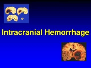
Intracranial Haemorrhages Intracerebral Extracerebral
Extradural (epidural) haemorrhage Arterial bleeding Bleeding from middle meningeal artery Following a hard blow to the head Blood collects between dura mata and skull Extradural haematoma is formed Haematoma biconvex in shape Brain compression and death if not evacuated
Subdural haematoma Venous bleeding Bleeding due to tearing of cerebral veins as they enter superior sagittal sinus Following a blow to the head that jerks the brain inside the cranial cavity Incident is usually long before and forgotten Blood collects between dura mata and arachnoid mater Subdural haematoma is sickle shaped
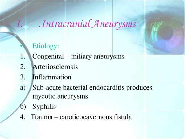
Subarachnoid haemorrhage Usually arterial bleeding Bleeding from internal carotid and circle of willis arteries Due to arterial aneurysms (ruptured due to high blood pressure) Blood collects in the subarachnoid space CSF gets mixed with blood----Xanthochromasia Severe headache and neck stiffness due to meningeal irritation
Sequence of events following head injury and raised intracranial pressure Initial concussion Lucid interval Drowsiness Pupils initially constrict then dilated and fixed Pulse initially may increase then reduce BP increases with reducing pulse (cushing reflex) Should monitor BP, pulse, respiration and pupils
Question • An individual received a fracture of the left temporal bone due to an accident. Subsequently, he developed extradural haemorrhage. Few hours later, he became drowsy and confused. An examination of his eyes showed a dilated left pupil that was non-reactive to light. His BP was high and pulse rate was reduced. • 1. Name the vessel involved in producing bleeding in this case. • 2. Explain the reasons for the following observations 2.1. Dilated, non-reactive pupil of left eye 2.2. Reduced pulse rate