Download
1 / 98
1.04k likes | 1.69k Views
Cardio pulmonary resuscitation . Cardiac arrest-abrupt cessation of cardiac pump function,which may be reversible Most victims of SCA demonstrate ventricular fibrillation (VF) at some point in their arrest.

E N D
Cardiac arrest-abrupt cessation of cardiac pump function,which may be reversible • Most victims of SCA demonstrate ventricular fibrillation (VF) at some point in their arrest. • Resuscitation is most successful if defibrillation is performed in about the first 5 minutes after collapse • High survival rates depends on a public trained in CPR and on well-organized public access defibrillation programs • Bystander CPR is performed in about only a third of witnessed arrests.
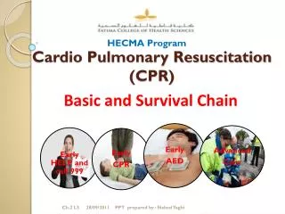
Adult Basic Life Support • BLS refers to maintaining airway patency and supporting breathing and the circulation, without the use of equipment other than a protective device • includes recognition of signs of sudden cardiac arrest (SCA), heart attack, stroke, and foreign-body airway obstruction (FBAO); cardiopulmonary resuscitation (CPR); and defibrillation with an automated external defibrillator (AED)
American Heart Association uses 4 links in a chain -the “Chain of Survival” to illustrate the important time sensitive actions for victims of VF SCA. • These links are • Early recognition of the emergency and activation of local emergency response system • Early bystander CPR: immediate CPR can double or triple the victim’s chance of survival from VF SCA. • Early delivery of a shock with a defibrillator: CPR plus defibrillation within 3 to 5 minutes of collapse can produce survival rates as high as 49% to 75% • Early advanced life support followed by postresuscitation care delivered by healthcare providers.
Adult BLS Sequence • Steps of BLS consist of a series of sequential assessments and actions, which are illustrated in the BLS algorithm
Open the Airway: Lay Rescuer • Head tilt– chin lift maneuver for both injured and noninjured victims. • The jaw thrust is no longer recommended for lay rescuers • Is difficult for lay rescuers to learn and perform • Is often not an effective way to open the airway • May cause spinal movement
Open the Airway: Healthcare Provider • Head tilt– chin lift maneuver to open the airway of a victim without evidence of head or neck trauma. • If cervical spine injury is suspected , open the airway using a jaw thrust without head extension • Use a head tilt– chin lift maneuver if the jaw thrust does not open the airway
Check Breathing • Look, listen, and feel for breathing. • Adequate breathing is not detected within 10 seconds, give 2 breaths • If you are a lay rescuer and you are unwilling or unable to give rescue breaths, begin chest compressions . • Treat the victim who has occasional gasps as if he or she is not breathing and give rescue breaths.
Give 2 rescue breaths, each over 1 second, with enough volume to produce visible chest rise. • recommendations for delivery of rescue breaths during cardiac arrest as follows • Deliver each rescue breath over 1 second • Give a sufficient tidal volume to produce visible chest rise • Avoid rapid or forceful breaths. • When an advanced airway (ie, endotracheal tube, Combitube, or LMA) is in place during 2-person CPR, ventilate at a rate of 8 to 10 breaths per minute without attempting to synchronize breaths between compressions
Mouth-to-Mouth Rescue Breathing • Open the victim’s airway • Pinch the victim’s nose • Create an airtight mouth-to-mouth seal. • Give 1 breath over 1 second • Take a “regular” (not a deep) breath • Give a second rescue breath over 1 second • Despite its safety, some may hesitate to give mouth-to-mouth rescue breathing and prefer to use a barrier device
Mouth-to-Nose Ventilation • Recommended if it is impossible to ventilate through the victim’s mouth,mouth cannot be opened or a mouth-to-mouth seal is difficult to achieve • Feasible, safe, and effective
Ventilation With Bag and Mask • Simultaneously open the airway with a jaw lift, hold the mask tightly against the patient’s face, and squeeze the bag. • The rescuer must also watch to be sure the chest rises with each breath • Most effective when provided by 2 trained and experienced rescuers • Rescuer delivers the breaths during pauses in compressions and delivers each breath over 1 second
Ventilation With an Advanced Airway • Advanced airway devices such as the LMA and the esophageal-tracheal combitube are currently within the scope of BLS practice in a number of regions • An advanced airway in place during CPR, 2 rescuers no longer deliver cycles of CPR . instead, • The compressing rescuer should give continuous chest compressions at a rate of 100 per minute without pauses for ventilation. • The rescuer delivering ventilation provides 8 to 10 breaths per minute. • The 2 rescuers should change compressor and ventilator roles approximately every 2 minutes • When multiple rescuers are present, they should rotate the compressor role about every 2 minutes.
Foreign-Body Airway Obstruction (Choking) • Recognition of airway obstruction is the key to successful outcome, it is important to distinguish this emergency • If mild obstruction is present and the victim is coughing forcefully, do not interfere with the patient’s spontaneous coughing and breathing efforts • Intervene if signs of severe obstruction develop • The cough becomes silent • Respiratory difficulty increases • Accompanied by stridor • The victim becomes unresponsive.
Chest thrusts, back slaps, and abdominal thrusts are feasible and effective for relieving severe FBAO in conscious (responsive) adults and children 1 year of age • Abdominal thrust- applied in rapid sequence until the obstruction is relieved • If abdominal thrusts are not effective, the rescuer may consider chest thrusts • Abdominal thrusts are not recommended for infants < 1 year of age because thrusts may cause injuries
If victim with FBAO becomes unresponsive, the rescuer should carefully support the patient to the ground, immediately activate EMS and then begin CPR. • Higher sustained airway pressures can be generated using the chest thrust rather than the abdominal thrust • Each time the airway is opened during CPR, the rescuer should look for an object in the victim’s mouth and remove it • Use a finger sweep only when the provider can see solid material obstructing the airway of an unresponsive patient (class indeterminate)
Rescue Breathing Without Chest Compressions • If an adult victim with palpable pulses requires support of ventilation, give rescue breaths at a rate of 10 to 12 breaths per minute • Reassess the pulse approximately every 2 minutes
Chest Compressions • Chest compressions consist of rhythmic applications of pressure over the lower half of the sternum • Blood flow generated by chest compressions delivers a small but critical amount of oxygen and substrate to the brain and myocardium • In VF chest compressions increase the likelihood that an attempted defibrillation will be successful • Chest compressions are especially important if the first shock is delivered 4 minutes after collapse
“Effective” chest compressions are essential for providing blood flow during CPR • to give “effective” chest compressions, “push hard and push fast.” Compress at a rate of about 100 compressions per minute, with a compression depth of 4 to 5 cm • Allow chest to recoil completely after each compression, and allow approximately equal compression and relaxation times. • Minimize interruptions in chest compressions.
Compression-Ventilation Ratio • A compression-ventilation ratio of 30:2 is recommended. • In infants and children 2 rescuers should use a ratio of 15:2 • Once an advanced airway is in place, 2 rescuers no longer deliver cycles of CPR • CPR prompt device may be useful in improving quality of CPR
Compression-Only CPR • Laypersons should be encouraged to do compression-only CPR if they are unable or unwilling to provide rescue breaths, although the best method of CPR is compressions coordinated with ventilations. • Survival rates were better with chest compressions only than with no CPR. • Rescue breathing is not essential during the first 5 minutes of adult CPR for VF. • If the airway is open, occasional gasps and passive chest recoil may provide some air exchange. • In addition, a low minute ventilation may be all that is necessary to maintain a normal ventilation-perfusion ratio during CPR.
Defibrillation • Early defibrillation is critical to survival from sudden cardiac arrest (SCA) for several reasons: • The most frequent initial rhythm in witnessed SCA is ventricular fibrillation (VF) • The probability of successful defibrillation diminishes rapidly over time • VF tends to deteriorate to asystole within a few minutes.
For every minute that passes between collapse and defibrillation, survival rates from witnessed VF SCA decrease 7% to 10% if no CPR is provided • When bystander CPR is provided, the decrease in survival rates is more gradual and averages 3% to 4% per minute • CPR can double or triple survival from witnessed SCA at most intervals to defibrillation. • CPR prolongs VF(ie, the window of time during which defibrillation can occur) and provides a small amount of blood flow that may maintain some oxygen and substrate delivery to the heart and brain
Shock First Versus CPR First • When any rescuer witnesses an out-of-hospital arrest and an AED is immediately available on-site, the rescuer should use the AED as soon as possible • Healthcare providers who treat cardiac arrest in hospitals and other facilities with AEDs on-site should provide immediate CPR and should use the AED/defibrillator as soon as it is available • When an out-of-hospital cardiac arrest is not witnessed by EMS personnel, they may give about 5 cycles of CPR before checking the ECG rhythm and attempting defibrillation particularly when call-to-response interval is > 5 min.
When VF is present for more than a few minutes, the myocardium is depleted of oxygen and metabolic substrates. A brief period of chest compressions can deliver oxygen and energy substrates, increasing the likelihood that a perfusing rhythm will return after defibrillation
1-Shock Protocol Versus 3-Shock Sequence • Interruption in chest compressions is associated with a decreased probability of conversion of VF in the 3-shock sequence. • Rhythm analysis of 3-shock sequence performed by commercially available AEDs revealed delays of up to 37 seconds between delivery of the first shock and delivery of the first post-shock compression • Animal studies documented harmful effects from interruptions to chest compressions • The rescuer should not delay resumption of chest compressions to recheck the rhythm or pulse. After 5 cycles (about 2 minutes) of CPR, the AED should then analyze the cardiac rhythm and deliver another shock if indicated.
The rescuer providing chest compressions should minimize interruptions in chest compressions for rhythm analysis and shock delivery • When 2 rescuers are present, the rescuer operating the AED should be prepared to deliver a shock as soon as the compressor removes his or her hands from the victim’s chest
Monophasic Waveform Defibrillators • deliver current of one polarity (ie,direction of current flow). • Biphasic Waveform Defibrillators • lower-energy biphasic waveform shocks have equivalent or higher success for termination of VF than monophasic waveform shocks delivering escalating energy (200 J, 300 J, 360 J) with successive shocks. • it is reasonable to use selected energies of 150 J to 200 J with a biphasic truncated exponential waveform or 120 J with a rectilinear biphasic waveform for the initial shock.
Automated External Defibrillators • AEDs are sophisticated, reliable computerized devices that use voice and visual prompts to guide lay rescuers and healthcare providers to safely defibrillate VF SCA • Lay rescuer AED programs to improve survival rates from out-of-hospital SCA • AEDs are of no value for arrest not caused by VF/pulseless VT, and they are not effective for treatment of nonshockable rhythms that may develop after termination of VF
Safe use of oxygen during defibrillation • Take off any oxygen mask/nasal cannulae & place them at least 1m away from the patient’s chest. • Leave the ventilation bag connected to the tracheal tube or other airway adjunct. • Alternatively, disconnect any bag-valve device from the tracheal tube (or other airway adjunct) and remove it at least 1m from the patient’s chest during defibrillation • Minimise the risk of sparks during defibrillation. • Self-adhesive defibrillation pads are less likely to cause sparks than manual paddles.
Electrode-patient interface • Transthoracic impedance (TTI)varies with body mass- approximately 70—80 Ω in adults • To reduce transthoracic impedance, the defibrillator operator should use conductive materials • Paddles should be well separated, and the paste or gel should not be smeared on the chest between the paddles • Defibrillation for patients with permanent pacemakers or ICDs, do not place the electrodes over or close to the device generator • Pacemakers and ICDs should be reevaluated after the patient receives a shock
Paddle electrodes and self-adhesive pad electrodes 8 to 12 cm in diameter perform well, although defibrillation success may be higher with electrodes 12 cm in diameter rather than with those 8 cm in diameter