Download
1 / 55
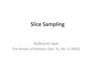
891 likes | 2.47k Views
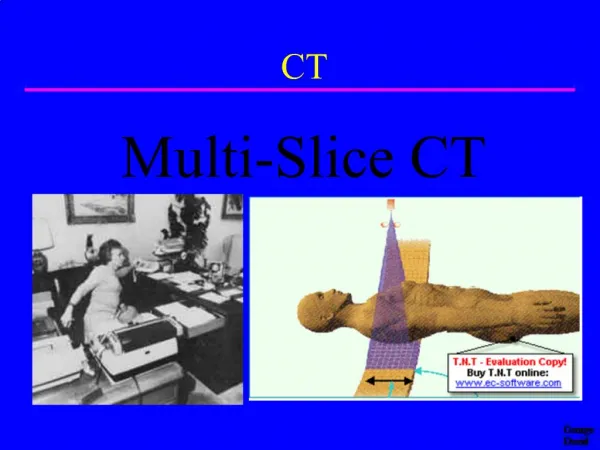
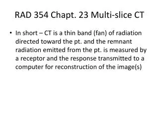
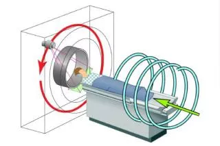
CT. Multi-Slice CT. Third Generation CT Single or Multislice. Z-axis orientation perpendicular to page. Patient. Single Slice Thickness Determined by Collimation. Z. Detector. Single-Slice Detectors. Many detectors rotate around patient Single row in z-direction

E N D
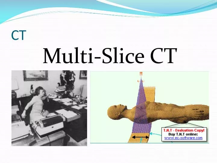
CT Multi-Slice CT
Third Generation CTSingle or Multislice Z-axis orientation perpendicular to page Patient
Single-Slice Detectors • Many detectors rotate around patient • Single row in z-direction • Slice thickness determined by collimation Z-Axis
Single Slice CT: Slice Thickness Collimated Beam Thickness Collimated Beam Thickness Thin slice Thick slice Z-Axis Z-Axis
Tube Multi-slice CT Post-collimator Detectors
Multislice CT • Multiple rows of detectors • Open collimators in “Z” direction 4 3 2 1 http://www.veterinary-imaging.com/images/MSS_CT.gif
Multi-slice CT • Developed in late 1990’s • Detector array segmented in z-direction • Simultaneous acquisition of multiple slices http://www.ctisus.com/gallery/images/multidetector/multislice_ct.jpg
Single Slice vs. Multislice Detector Collimated Beam Thickness Multislice detector Single slice detector Z-Axis
Multi-Slice Detectors • Many detectors going around patient • Many detector rows in z-direction • Slice thickness determined by • Collimation • electronic detector selection “Z” Direction Multi Single
Multi-slice CT • Size & distribution of detectors in non-axial direction similar to previous CT’s • Similar spatial & contrast resolution
Distribution of detectors in axial direction varies with manufacturer All detectors same width Variable widths “Z”Direction
Multi-slice CTUniform Detector Thickness “z” direction • Multiple detectors in axial direction • Size must accommodate thinnest slice • Detector signals can be used • Individually • In groups 1 2 3 4 Four thin slices 4 1 2 3 Four thicker slices
Detectors vs. Channels • # Physical Detectors not necessarily equal to # of possible Slices • Maximum # slices limited by Digital Acquisition System (DAS) channels • Electronic counters • Imaging speed bottleneck • How fast data can be received fromdetector arrays
Detectors vs. Channels Example • 16 detectors • 4 channels
Detectors vs. Channels4 X 1.25 mm • Beam collimated to 4 detector rows • 1 detector row per DAS channel Effective Detector
Detectors vs. Channels4 X 2.5 mm • Beam collimated to 8 detector rows • 2 detector rows per DAS channel Effective Detector
Detectors vs. Channels4 X 3.75 mm • Beam collimated to 12 detector rows • 3 detector rows per DAS channel Effective Detector
Detectors vs. Channels4 X 5 mm • Beam collimated to 16 detector rows • 4 detector rows per DAS channel Effective Detector
Capture Efficiency • Fraction of detector area that is active detector
Equal-width Detectors Disadvantage • Many gaps • Gaps are dead space • Reduce capture efficiency
Multi-slice CT“Adaptive Array Detectors” “z” direction • Some scanners use detectors of various widths • Post-collimators used to partially block wider elements for thinner slices 3 1 2 Three thicker slices Post-collimators 2 1 3 4 Four thinner slices
Variable Width Detectors • Center detectors thinner • Thicker detectors can function as thinner ones using collimation • Thinner detectors can function a thicker one by combining signals
Single Slice Pitch Definition table motion during one rotation Slice Pitch = --------------------------------------- slice thickness
Beam thickness Beam PitchDefined only for Multi-slice scanners table motion during one rotation Beam Pitch = --------------------------------------- Beam thickness
Beam PitchDefined only for Multi-slice scanners Beam Pitch = 1 Beam Pitch > 1
Example • 5 mm slices • 4 simultaneous slices • Beam pitch = 1 • 1 revolution / sec. • Beam thickness? • Table speed? table motion during one rotation Beam Pitch = --------------------------------------- Beam thickness
Beam Thickness • 5 mm slices • 4 simultaneous slices • Beam pitch = 1 • 1 revolution / sec. table motion during one rotation Beam Pitch = --------------------------------------- Beam thickness Beam Thickness = 5 X 4 = 20 mm
Table Speed • 5 mm slices • 4 simultaneous slices • Beam pitch = 1 • 1 revolution / sec. • 20 mm beam thickness table motion during one rotation Beam Pitch = --------------------------------------- Beam thickness Table speed = 20 mm rotation (1 sec) = 20 mm / sec
Tube Slice Thickness Defined at Rotational Center Rotational Center
Detector Field must be Larger than Slice Thickness at Rotational Center Diverging Beam Rotational Center
Beam Divergence More of a Problem for Multi-Slice • Rays diverge • No longer essentially parallel • Leads to Cone Angle Artifact • Significant for 16, 32, 64 … data channels • Requires use of special reconstruction algorithms to compensate
Multislice CT Doses • Can be 10-30% higher than for single slice units (ICRP #47) • Cause • Divergent beam • Other considerations • Tendency to cover more volume (anatomy) • Better availability of equipment
Other Reasons for High CT Doses • Repeat Exams • No adjustment of technique factors for different size patients • No adjustment for different areas of body
Multislice CT Advantage? Speed!
Speed = Power • Speed enables new applications
Multi-slice CT ImagingClinical Advantages • Thinner slices for improved z-direction resolution • Same acquisition in shorter time • Scan larger volumes in same time
Multi-slice CT ImagingClinical Advantages • Thinner slices • Improvement in CTA of neck, aorta, renal vessels • Better reconstructions • Sagittal, coronal, oblique • 3-D • Fundamental Trade-off • “z” axis resolution vs. image noise
Multi-slice CT ImagingClinical Advantages • Improved x-ray tube utilization • Reduced x-ray tube loading • 4 slices acquired with same tube loading previously used for 1 • Less need to pause of tube cooling • Reduced wear & tear • Other anticipated benefits • CT endoscopy • Diagnosis of pulmonary embolism
Multi-slice CT ImagingClinical Advantage: Angiography • Simplifies contrast bolus timing • Continuous observation of target vessel • Can reduce amount of contrast required • Coverage from aorta to lower extremities • Runoff CTA ~ 13% of all CT procedures
Continuous CT ImagingInterventional Procedures • Biopsy & drainage • Neuro • Chest • Abdominal • Spine • Catheter and tube placement • Helps operator avoid critical structures near path of biopsy needle • Better visualizing of moving structures • Respiration • Functional CT • Brain perfusion
Multi-Slice Compared to Single-slice helical • Much Faster • No significant image quality differences • Equivalent Patient Dose • Ref: • Willi Kalender, Ph.DInstitute of Medical PhysicsUniversity of Erlanger, Germany
Organ Coverage Time Depends On • Beam Pitch • Rotation Time • Detector Acquisition Length
64 Slice CT Typical Coverage Times • Heart & coronary arteries / brain / lungs • 5 seconds • Whole body coverage for blood clot search • 30 seconds Philips
64-Slice Commercial Cardiac CT www.impactscan.org