Download

1 / 24
240 likes | 340 Views
Blank Pics for Head, Neck and Face Muscles. BIO 238. OCCIPITAL BONE. Lambdoid suture. PARIETAL BONE (right). PARIETAL BONE (left). Sagittal suture. Coronal suture. FRONTAL BONE. ZYGOMATIC BONE. NASAL BONES. Superior view. Coronal suture. FRONTAL BONE. PARIETAL BONE.

E N D
OCCIPITAL BONE Lambdoid suture PARIETAL BONE (right) PARIETAL BONE (left) Sagittal suture Coronal suture FRONTAL BONE ZYGOMATIC BONE NASAL BONES Superior view
Coronal suture FRONTAL BONE PARIETAL BONE SPHENOID Supra-orbital foramen Squamous suture TEMPORAL BONE NASAL BONE LACRIMAL BONE Squamous part of temporal bone ETHMOID Lambdoid suture Infra-orbital foramen OCCIPITAL BONE MAXILLA External acoustic meatus ZYGOMATIC BONE Mastoid process Styloid process MANDIBLE Zygomatic process of temporal bone Mental foramen Zygomatic arch Temporal process of zygomatic bone Mental protuberance Lateral view
Coronal suture PARIETAL BONE FRONTAL BONE SPHENOID TEMPORAL BONE Supra-orbital foramen ETHMOID Optic canal PALATINE BONE Superior orbital fissure LACRIMAL BONE Inferior orbital fissure Temporal process of zygomatic bone ZYGOMATIC BONE Mastoid process of temporal bone Infra-orbital foramen NASAL BONE Middle nasal concha (part of ethmoid) MAXILLA Perpendicular plate of ethmoid INFERIOR NASAL CONCHA Bony nasal septum VOMER MANDIBLE Mental protuberance Anterior view Mental foramen
FRONTAL BONE MAXILLA ZYGOMATIC BONE VOMER SPHENOID PALATINE BONE Foreman ovale Zygomatic arch Medial and lateral pterygoid processes Styloid process Mandibular fossa Foramen lacerum Carotid canal External acoustic meatus TEMPORAL BONE Mastoid process Jugular foramen Stylomastoid foramen Lambdoid suture Occipital condyle OCCIPITAL BONE Foramen magnum External occipital protuberance Inferior view
FRONTAL BONE Crista galli ETHMOID Cribriform plate Sella turcica Foramen rotundum SPHENOID Foramen lacerum Foramen ovale Foramen spinosum TEMPORAL BONE Carotid canal Internal acoustic meatus Foramen magnum PARIETAL BONE Jugular foramen Internal occipital crest Hypoglossal canal OCCIPITAL BONE Superior view of a horizontal section through the skull, showing the floor of the cranial cavity.
Hypoglossal canal Occipital condyle Foramen magnum External occipital crest Inferior nuchal line Superior nuchal line External occipital protuberance Occipital bone, inferior view
Superior temporal line Inferior temporal line Right parietal bone, lateral view
Frontal (metopic) suture Frontal squama Superior temporal line Supra-orbital notch Supra-orbital margin Anterior surface
Squamous part Mandibular fossa External acoustic meatus Zygomatic process Styloid process Mastoid process Lateral view of the right temporal bone
Sphenoidal sinus Lesser wing Superior orbital fissure Orbital surface Greater wing Body Foramen rotundum Pterygoid process Pterygoid plates Anterior surface
Orbital rim Infra-orbital foramen Anterior nasal spine Alveolar process Zygomatic process An anterolateral view of the right maxilla.
Supra-orbital foramen NASAL BONE SPHENOID TEMPORAL BONE Zygomaticofacial foramen ZYGOMATIC BONE Infra-orbital foramen MAXILLA Perpendicular plate of ethmoid VOMER Bony nasal septum
Articular surface for temporomandibular joint Coronoid process Teeth Mandibular notch Alveolar process Head Ramus Mental protuberance Body Condylar process Mental foramen Angle A lateral and slightly superior view of the mandible
Greater horn Lesser horn Body An anterior view of the hyoid bone
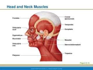
Epicranial aponeurosis Temporoparietalis (cut and reflected) Frontal belly of occipitofrontalis Temporalis Procerus Orbicularis oculi Nasalis Occipital belly of occipitofrontalis Levator labii superioris Zygomaticus minor Masseter Levator anguli oris Buccinator Zygomaticus major Sternocleidomastoid Mentalis (cut) Orbicularis oris Trapezius Depressor labii inferioris Depressor anguli oris Omohyoid Platysma (cut and reflected) Lateral view
Scalenes Anterior Middle Posterior Anterior view, cervical region
Superior temporal line Temporalis Capsule of temporomandibular joint Zygomatic arch Masseter Lateral view. The temporalis muscle passes medial to the zygomatic arch to insert on the coronoid process of the mandible. The masseter inserts on the angle and lateral surface of the mandible.
Mylohyoid (cut and reflected) Mandible Mylohyoid Geniohyoid Digastric Stylohyoid Anterior belly Hyoid bone Posterior belly Thyrohyoid Sternocleidomastoid (cut) Thyroid cartilage of larynx Omohyoid Sternothyroid Superior belly Inferior belly Sternohyoid Clavicle Sternocleidomastoid Clavicular head Cut heads of sternocleidomastoid Sternal head Sternum Anterior view
Epicranial aponeurosis Frontal belly of occipitofrontalis Temporoparietalis (cut and reflected) Corrugator supercilii Temporalis Temporalis (temporoparietalis removed) Orbicularis oculi Procerus Nasalis Levator labii superioris Zygomaticus minor Levator anguli oris Zygomaticus major Masseter Orbicularis oris Buccinator Risorius Platysma Depressor anguli oris Depressor labii inferioris Sternal head of sternocleidomastoid Mentalis (cut) Thyroid cartilage of the larynx Clavicular head of sternocleidomastoid Trapezius Clavicle Platysma (cut and reflected) Anterior view
Epicranial aponeurosis Temporoparietalis (cut and reflected) Frontal belly of occipitofrontalis Temporalis Procerus Orbicularis oculi Nasalis Occipital belly of occipitofrontalis Levator labii superioris Zygomaticus minor Masseter Levator anguli oris Buccinator Zygomaticus major Sternocleidomastoid Mentalis (cut) Orbicularis oris Trapezius Depressor labii inferioris Depressor anguli oris Omohyoid Platysma (cut and reflected) Lateral view
Lateral pterygoid Medial pterygoid Cut edge of mandible Lateral view, pterygoid muscles exposed. The location and orientation of the pterygoid muscles can be seen after the overlying muscles, along with a portion of the mandible, are removed.
Erector Spinae, Deep Layer Intervertebral Muscles, Posterior View Spinal Flexors Quadratus lumborum Rotatores Intertransversarii Flexors of the Anterior Cervical and Thoracic Spine Longus capitis Spinous process of vertebra Thoracodorsal fascia Posterior view Longus colli Interspinales Transverse process of vertebra