Download
1 / 21
220 likes | 352 Views
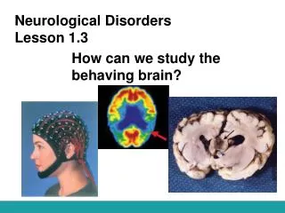
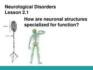
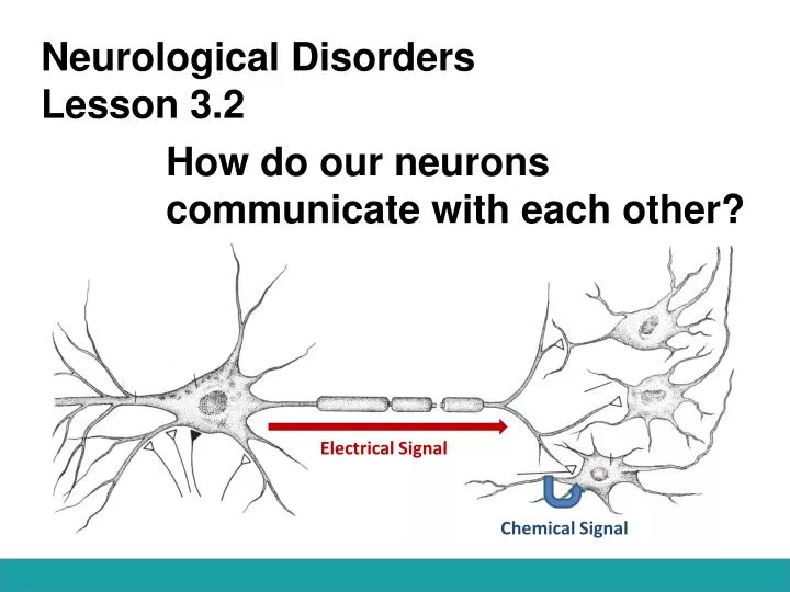
Neurological Disorders Lesson 3.2. How do our neurons communicate with each other?. Electrical Signal. Chemical Signal. Do Now:. Sleeping Beauty just pricked her finger and is feeling a lot of pain. Model the neurons involved in Sleeping Beauty sensing this pain.

E N D
Neurological DisordersLesson 3.2 How do our neurons communicate with each other? Electrical Signal Chemical Signal
Do Now: • Sleeping Beauty just pricked her finger and is feeling a lot of pain. Model the neurons involved in Sleeping Beauty sensing this pain. • In order for one neuron in this pathway to send information to the next, how would you change the electrical signal of the axon into a chemical signal at the synapse ?
Pain Pathway Sensory neuron Projection neuron Motor neuron Interneuron
Converting an Electrical Signal to Chemical Signal Electrical Signal Chemical Signal
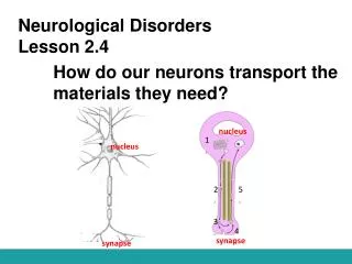
Synaptic Transmission Electrical Signal Neurotransmitter
Synaptic Transmission Electrical Signal Neurotransmitter
The Stage: Presynaptic cell Synapse Postsynaptic Cell
The Characters: Voltage-gated Ca2+ channels Synaptic vesicles Neurotransmitters (NT) Action Potential Receptors Ca2+ sensitive proteins Reuptake Transporters
How do the characters work together to complete synaptic transmission? Card Sort Activity
The Play: 1. Action Potential
The Play: 2. Voltage-gated Ca2+ channels open. 1. Action Potential
The Play: 2. Voltage-gated Ca2+ channels open. 3. Ca2+ flows into cell Ca2+ 1. Action Potential
The Play: 2. Voltage-gated Ca2+ channels open. 4. Ca2+ sensitive proteins fuse synaptic vesicles to membrane. 3. Ca2+ flows into cell Ca2+ 1. Action Potential
The Play: 2. Voltage-gated Ca2+ channels open. 4. Ca2+ sensitive proteins fuse synaptic vesicles to membrane. 3. Ca2+ flows into cell 5. NTs are released into synaptic cleft Ca2+ 1. Action Potential
The Play: 2. Voltage-gated Ca2+ channels open. 4. Ca2+ sensitive proteins fuse synaptic vesicles to membrane. 6. NTs bind to postsynaptic receptors. 3. Ca2+ flows into cell 5. NTs are released into synaptic cleft Ca2+ 1. Action Potential
The Play: 2. Voltage-gated Ca2+ channels open. 4. Ca2+ sensitive proteins fuse synaptic vesicles to membrane. 6. NTs bind to postsynaptic receptors. 3. Ca2+ flows into cell 5. NTs are released into synaptic cleft Ca2+ 1. Action Potential 7. Ion channels open on postsynaptic membrane, allowing ions to flow into cell.
The Play: 2. Voltage-gated Ca2+ channels open. 4. Ca2+ sensitive proteins fuse synaptic vesicles to membrane. 6. NTs bind to postsynaptic receptors. 3. Ca2+ flows into cell 5. NTs are released into synaptic cleft Ca2+ 1. Action Potential 7. Ion channels open on postsynaptic membrane, allowing ions to flow into cell. 8. Excess NTs are degraded by enzymes or pumped back into presynaptic cell.
What would happen if… You took a drug that destroyed the Ca2+ sensitive proteins that fuse synaptic vesicles to the membrane??? You wouldn’t be able to release synaptic vesicles.
That’s How Botox Works! Before After Botox destroys the proteins that fuse synaptic vesicles with the membrane. By stopping vesicle release, Botox prevents muscle contraction which prevents wrinkles!
Synaptic Transmission 2. Voltage-gated Ca2+ channels open. Ca2+ flows into cell 3. Ca2+ sensitive proteins fuse synaptic vesicles to membrane, releasing NTs into synaptic cleft 4. NTs bind to postsynaptic receptors. Ca2+ 1. Action Potential 5. Ion channels open on postsynaptic membrane, allowing ions to flow into cell. 6. Excess NTs are degraded by enzymes or pumped back into presynaptic cell.
Does it matter which ions flow into the postsynaptic cell? Sodium (Na+) Calcium (Ca2+) Chloride (Cl-) Positive Positive Negative