Download
1 / 41
430 likes | 625 Views
HOW TO READ ELECTROCARDIOGRAPHY. SYARIF HIDAYATULLAH STATE ISLAMIC UNIVERSITY (UIN), JAKARTA. Dr. Yasmin Tadjoedin , Sp.JP. ECG INTERPRETATION. RATE RHYTHM AXIS HIPERTROPHIC SIGNS MYOCARDIAL INFARCTION ARRHYTHMIA. Rhytm QRS Rate QRS Axis P wave PR Interval QRS Duration

E N D
HOW TO READ ELECTROCARDIOGRAPHY SYARIF HIDAYATULLAH STATE ISLAMIC UNIVERSITY (UIN), JAKARTA Dr. YasminTadjoedin, Sp.JP
ECG INTERPRETATION • RATE • RHYTHM • AXIS • HIPERTROPHIC SIGNS • MYOCARDIAL INFARCTION • ARRHYTHMIA
Rhytm QRS Rate QRS Axis P wave PR Interval QRS Duration QRS Morphology ST Segmen Deviation T wave morphology U wave Others (LVH,LV Strain,BBB, QT interval) ECG conclusion STEPS IN ECG READING : NORMAL VALUES : PR Interval 0,12’’ - 0,20’’ QRS Duration 0,04’’ - 0,12’’ Normal Axis - 300 - + 1100
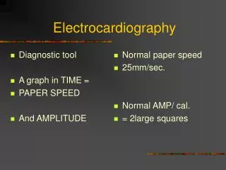
1. RATE • Normal heart rate : 60 – 100 x/minutes • > 100 x/minutes : Sinus Tachycardia • < 60 x/minutes : Sinus Bradicardia • Determination heart rate (normal paper speed 25 mm/s): • 300 • Count number of large square (bold boxes in one R – R’ interval) • 1500 • Count number of small square in one R – R’ intervals • Number of QRS complex in 6 seconds, multiply by 10
2. RHYTHM Normal cardiac rhythm : SINUS rhythm • Sinus rhythm characteristics : • Rate 60-100 bpm • Constant R – R interval • Negative P wave in aVR and positive di II • P wave is always followed by QRS complex
4. HYPERTROPHIC SIGNS LEAD II
5. MYOCARDIAL INFARCTION • Ischemia • Injury • Necrosis
WHAT’S WRONG?? Lead Error: V1 and V3 are Transposed! In this normal 12-lead ECG the V1 and V3 chest electrodes are interchanged. Experienced ECG interpreters should be able to spot this lead placement error.