Download
1 / 3
30 likes | 95 Views
Nanomagnetic Structures for Atomic Resolution MRI. Mladen Barbic, CSU Long Beach, CAREER DMR 0349319.

E N D
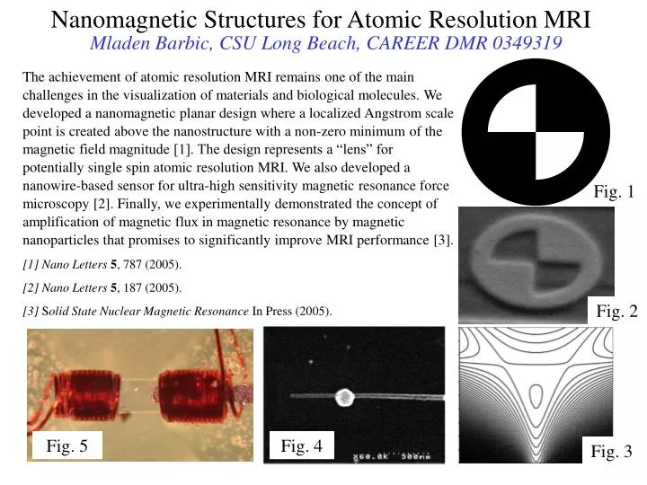
Nanomagnetic Structures for Atomic Resolution MRI Mladen Barbic, CSU Long Beach, CAREER DMR 0349319 The achievement of atomic resolution MRI remains one of the main challenges in the visualization of materials and biological molecules. We developed a nanomagnetic planar design where a localized Angstrom scale point is created above the nanostructure with a non-zero minimum of the magnetic field magnitude [1]. The design represents a “lens” for potentially single spin atomic resolution MRI. We also developed a nanowire-based sensor for ultra-high sensitivity magnetic resonance force microscopy [2]. Finally, we experimentally demonstrated the concept of amplification of magnetic flux in magnetic resonance by magnetic nanoparticles that promises to significantly improve MRI performance [3]. [1] Nano Letters5, 787 (2005). [2] Nano Letters5, 187 (2005). [3] Solid State Nuclear Magnetic Resonance In Press (2005). Fig. 1 Fig. 2 Fig. 5 Fig. 4 Fig. 3
Nanomagnetic Structures for Atomic Resolution MRI Mladen Barbic, CSU Long Beach, CAREER DMR 0349319 Societal Impact: Our laboratory serves the urban, ethnically and culturally diverse student community of southern California. We focus on providing the opportunity to undergraduate and masters students to gain valuable knowledge, research experience, and exposure to the forefronts of nanotechnology. By learning to construct novel state-of-the-art scientific instrumentation and devices, the students from traditionally underrepresented groups can actively contribute to the academic advancements in nanotechnology. Education: Masters Degree Students: Mary Brady studied the angular dependence of magnetic nanoparticle amplification and successfully completed and defended her Master Degree thesis based on NSF support. Michael Hetman is studying the MRI “lens” design optimization towards atomic resolution MRI for his Masters Thesis project. Gabriel Lucero is investigating novel magnetic sensors for improved MRI detection under the NSF support of his Masters Degree. Undergraduate Students: Robert Lugo is developing micro-coil designs for ultra-high sensitivity magnetic measurements under NSF support and will attend medical school next year. BJ Gorman developed the ultra-high sensitivity magnetometer under the NSF support and graduated with a BS in Physics. He currently works at a defense company in the Los Angeles area. Fig.6
Nanomagnetic Structures for Atomic Resolution MRI Mladen Barbic, CSU Long Beach, CAREER DMR 0349319 Fig. 1 Magnetic resonance microscopy “lens” design that consists of a 10nm thick perpendicular anisotropic magnetic material disk with two inside quarter-circle diagonally opposed cuts. Outside radius is 60nm and inside radius is 40nm. A bias field opposite to the magnetization direction is also required for obtaining a localized magnetic field magnitude minimum. Fig. 2. Planar nanostructure fabricated by using the Focused Ion Beam lithography. Fig. 3. Contours of constant magnitude of the magnetic field above the “lens” structure. The “focus” or the localized magnetic field magnitude minimum is located at z=23.8(nm) above the plane. Only the spins within the central contour satisfy the magnetic resonance condition, and would be detected. Fig. 4. Scanning Electron Microscopy image of the composite nanowire-based Magnetic Resonance Force Microscopy sensor. Magnetic nickel section is on the left, followed by the silver nano-reflector, and completed by the platinum nanowire-based cantilever structure. Fig. 5. The experimental arrangement for simulating magnetic resonance amplification by magnetic nanoparticles. Two micro-coils are wound on a hollow rectangular capillary tube and distanced by approximately 250μm. Approximately 50μm wide, 12.5μm thick tape strip (with 5.5μm thick layer of magnetic nanoparticles) is cut along the nanoparticle long axis direction and forms the core of one of the micro-coils. The set-up is placed between the poles of an electro-magnet so that the tunable DC magnetic field is applied perpendicular to the micro-coils and nanoparticle easy axis. The micro-coil on the left simulates the spin sample as the source of time-dependent magnetic fields that are small and perpendicular to the DC magnetic field, while the nanoparticle-filled micro-coil on the right serves as the magnetic flux detector. Fig. 6. Detected AC micro-coil voltage at 3MHz reveals a peak at the DC field of ~1,250 (Gauss), indicating the singularity in the reversible transverse susceptibility RT of single magnetic domain nanoparticles. The detected voltage from the micro-coil detector with the nanoparticles removed is also shown on the graph for comparison. The magnetic core material amplifies the AC magnetic flux through the inductive coil at an appreciable value of the perpendicular DC magnetic field, as required for magnetic resonance settings.