Download
1 / 14
160 likes | 343 Views
BioE 498/598 DP 04-16-2014 Optical Coherence Tomography. Introduction of OCT. James G. Fujimoto, 1991 What is OCT: diagnostic medical imaging technology Why OCT: better diagnose and treat disease Main application areas: heart disease, cancer, ocular disease.

E N D
Introduction of OCT • James G. Fujimoto, 1991 • What is OCT: diagnostic medical imaging technology • Why OCT: better diagnose and treat disease • Main application areas: heart disease, cancer, ocular disease In situ imaging of tissue microstructure with a resolution approaching that of histology, but without the need for tissue excision and processing
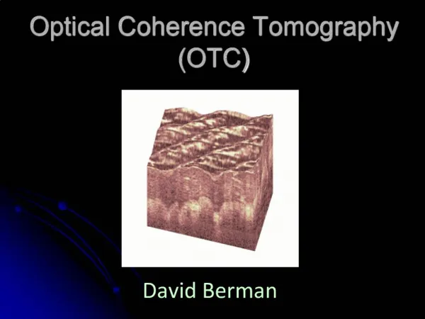
What is OCT? • OCT use low-coherence interferometry to produce a two or three dimensional image of optical scattering from internal tissue microstructures. • Michelson interferometer is used to perform low-coherence interferometry • OCT measures intensity of reflected infrared light. • Based on measurements of the reflected light from tissue discontinuities • e.g. the epidermis-dermis junction. • Based on interferometry • involves interference between the reflected light and the reference beam. http://biophotonics.illinois.edu/pubs/biophotonics_current/OCTareviewfrombenchtobedside.pdf
Resolution (log) 1 mm Ultrasound 100 mm 10 mm Confocalmicroscopy 1 mm Penetration depth (log) 10 cm 1 mm 1 cm OCT vs. standard imaging Standardclinical Highfrequency OCT
Fundamental OCT Schematic Broadband source Fiber-optic beamsplitter Tissue Detector Scanning reference mirror Computer Bandpass filter Amplifier
OCT in Nontransparent Tissue B arterial layers A epiglottis C atherosclerotic plaques
Advantage of OCT • Broad dynamic range, • High resolution • Rapid data acquisition rate, • Small inexpensive catheter/endoscope design • Compact portable structure (fiber optically based, making possible the development of small catheters and endoscopes) • The frame rate for OCT systems are four to eight frames per second.(assume an image size of 256 by 512 pixels.)
OCT application Esophagus & epithelium & early cancer Vulnerable plaque B Reduce Biopsy Hazardous Prostate Applied in guiding microsurgical procedure A Reduce High False-Negative Rates
Polypyrrole Nanoparticles relatively low concentration of biocompatible PPy nanoparticles can improve the OCT Image contrast of an intralipid tissue phantom.
FIBEROPTIC PROBE INTEROMETER ELECTRONICS AND OPTICS +COMPUTER DISPLAY AND KEYBOARD Nowadays and future equipment • Low-coherence Superluminescent diode:800 –1300 nm center waveength and several milliwatts power. Not available for salePending 510(k)
Limitation • Penetration: 2-3mm Ideal: 4mm • Resolution : catheter/endoscope based image: 10μm, noncatheter: 4 μm, 1. femtosecond laser is expensive (1 μm) 2. transverse resolution needs to be similar to axial resolution, below 10 μm need short confocal parameter which results in the focus falling off rapidly. • Acquisition rate: <10franes/second • Lack of large-scale clinical trials