Download
1 / 33
340 likes | 379 Views
Muscular Dystrophies. What is Muscular Dystrophy? (MD). MD is an inherited muscle disease with many different forms. In most cases muscle progressively become weaker. Some types of MD affect voluntary muscles such as the heart.

E N D
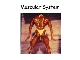
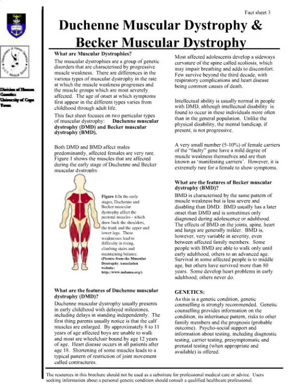
What is Muscular Dystrophy?(MD) • MD is an inherited muscle disease with many different forms. • In most cases muscle progressively become weaker. • Some types of MD affect voluntary muscles such as the heart. • Muscular Dystrophy: group of genetic disorders that are characterized by progressive loss of muscle integrity, wasting, and weakness. Characterized by degeneration and regeneration of muscle fibers (in contrast with static or structural myopathies) • Muscular Dystrophy Association • Covers all muscular dystrophies and myopathies • Multisystem diseases : ALS or Friedreich Ataxia • Neuropathy : HSMN, CMTD
Muscular dystrophy refers to a group of genetic, hereditarymuscle diseases that cause progressive muscle weakness. • Muscular dystrophies are characterized by progressive skeletal muscle weakness, defects in muscle proteins, and the death of muscle cells and tissue
Symptoms • Lack of coordination • Muscle weakness • Progressive crippling • Loss of mobility
General Diagnostic Testing • Creatine kinase : • greatly elevated (50 times normal) • Increased in DMD, BMD, polymyositis, and rhabdomyolysis • Nonspecific if mildly elevated 2-3x normal • Lower late in MD course due to severely reduced muscle mass • Not helpful for carrier detection
Muscle biopsy • Dystrophic changes include necrosis, degeneration, regeneration, fibrosis and fatty infiltration, sometimes mild inflammation • Specific diseases may have inflammation, intracellular vacuoles, rods, and other inclusions on biopsy • Biochemical muscle protein analysis • Useful for specific identified protein that is missing and many specific mutations may cause the same deficiency • Immunohistochemical protein staining • Western blot – quantitates percent of normal protein present
Genetic analysis • PCR for specific known defects • Southern blot for nucleotide repeats • Electromyography • Useful if diagnosis not clear (biopsy has mixed features) • Differentiates neuropathic vs. myopathic • Characteristic myotonic discharges in adults with myotonia – “dive bomber” sound • Perform after the CK
CLASSIFICATION X-linked muscular dystrophy • Duchenne muscular dystrophy • Becker muscular dystrophy • Emery-Dreifuss syndrome
CLASSIFICATION Autosomal recessive muscular dystrophy • Limb-girdle muscular dystrophy Autosomal dominant muscular dystrophy • Fascioscapulohumeral muscular dystrophy
Differential Patterns of Initial Muscle Weakness in MD Congenital MD Duchenne MD OPMD Oculopharyngeal MD Emery Dreifuss MD Limb Girdle MD FSHD Facio- Scapulo- humeral
Congenital Muscular Dystrophy • Presentation: neonatal onset of severe weakness, delayed motor milestones, contractures • Merosin negative/CMD A1 • White matter hypodensities on brain scan but normal mental capacity • Diagnosis by muscle biopsy immunohistochemistry showing loss of α2-laminin (AR-chromosome 6q22-23)
Duchenne Muscular Dystrophy • Presentation: 3-5 y/o with frequent falls, slow running • Prevalence of 1:3500 • Etiology • single gene defect (65% deletions, 7-10% duplications, 25% point mutations, small deletions or insertions) • 1/3 new mutation • 2/3 family history • Xp21.2 region • absent dystrophin
Duchenne Muscular Dystrophy • Clinical Manifestations • Onset : age 3-6 years • Progressive weakness • Pseudohypertrophy of calf muscles • Spinal deformity • Cardiomyopathy • Respiratory • 30% mild to moderate MR
Pseudohypertrhophy of calf muscle, • Tip toe gait • forward tilt of pelvis, compensatory lordosis
Duchenne Muscular Dystrophy • Natural History • Progress slowly and continuously • muscle weakness • lower --> upper extremities • unable to ambulate: 10 year (7-12) • death from pulmonary/ cardiac failure: 2-3rd decade
Clinical Signs (Gower’s Sign) Family history (pedigree analysis) Increase CPK (200x) DNA mutation analysis (65%) or haplotype analysis) Myopathic change in EMGBx: m. degeneration Muscle biopsy and Immunoblotting: Absence dystrophin (if geneticist can’t find the mutation !!) Prenatal diagnosis is available Duchenne Muscular DystrophyDiagnosis
Becker Muscular Dystrophy • Slowly progressive form with same gene affected as Duchenne MD • Etiology • single gene defect • short arm X chromosome • altered size & decreased amount of dystrophin • Muscle biopsy immunostaining for dystrophin with patchy staining • Disorder of function or decreased amount of dystrophin rather than absence of the protein
Emery-Dreifuss Muscular Dystrophy • Presentation: Thisdisorder is characterizedby a triad • Earlycontractures of theAchillestendon, elbowsandposteriorcervicalmuscles • Slowlyprogressivemusclewastingandweaknesswith a humeroperonealdistribution • Cardiomyopathyarises , whichusuallypresents as cardiacconductiondefects. • Genetics • X-linked type affects emerin (STA gene at chromosome Xq28) • Diagnose by protein analysis of leukocytes or skin fibroblasts • DNA testing available • AD affects lamin A or lamin C (chromosome 1q21)
Limb Girdle Muscular Dystrophy • Presentation: variable age of onset with weakness and wasting of the limb-girdle • 15 genetically different types (genetical and clinical heterogenic) • AD forms are rare but more less severe than AR forms • Several of these disorders are associated with clinically significant cardiac involvment
FascioScapularHumeral Muscular Dystrophy (AD) • Presentation: • Facial and shoulder girdle are first affected muscle group • Later foot extensors and pelvic girdle muscles become involved • The heart is not implicated in most cases. • mild high pitched hearing loss, retinal abnormalities, mental retardation in early onset • Genetics/Testing • Southern blot testing available (chromosome 4q35) for decrease in repeats normally present • Muscle biopsy may show lymphocytic infiltrates
Oculopharyngeal Muscular Dystrophy • Presentation: • mid-adult with ptosis, facial muscle weakness with difficulty swallowing, proximal muscle weakness, may have extraocular muscle weakness, more common in French-Canadian and Hispanic population • Genetics • Affects poly A binding protein 2 (PABP2) by expansion of a GCG repeat without anticipation seen – Southern blot (chromosome 14q11-13)
Myotonic Dystrophy • Presentation – adult with multiple systems affected • Primarily distal and facial weakness • Facial features: frontal balding in men, ptosis, low-set ears, hatchet jaw, dysarthria, swan neck, ^ shaped upper lip • Myotonia: worse in cold weather, after age 20 • Heart: conduction block – evaluate syncope • Smooth muscle: constipation, care with swallowing, gallstones, problems with childbirth, BP lability • Brain: learning disabilities, increased sleep requirement • Ophthalmology: cataracts • Endocrine: insulin resistance, hypothyroidism, testicular atrophy
Huntington Disease • Presentation • Mood Swings • Impaired cognitive functions • Chorea • Huntington’s Disease is an Autosomal Dominant“Tri-nucleotide Repeat” Disorder caused by a mutation of a gene on the 4th chromosome which is responsible for producing the protein Huntingtin, that creates excess copies of the CAG codon which genetically program the degeneration of the neurons of the brain. • Age of onset is found generally in adults around the age of 40 • The earliest onset of Huntington’s ever documented was a two year old boy. • The symptoms of HD can also develop at 55 or later, in which case it is harder to recognize.
The number of CAG codons varies and so does the severity of the disease • >40 repeats you develop HD, children 50% chance of developing disease • 36-39 repeats “Grey Zone” May develop HD, children may or may not develop HD • 29-35 repeats the individual will not develop HD, children may • <29 repeats, the individual will not develop HD, children will not develop HD
Treatment • There is no cure for MD • Medications that are prescribed for MD patients • Steroids • Braces for support • Mobility chairs • Surgery is also an option to release contractures