Download
1 / 42
420 likes | 475 Views
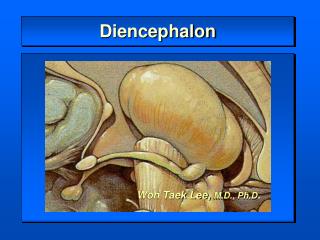
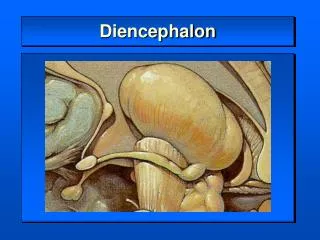
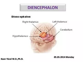
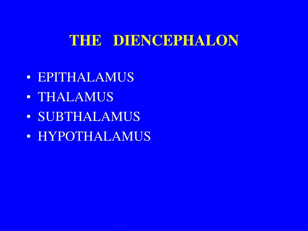
THE DIENCEPHALON. EPITHALAMUS THALAMUS SUBTHALAMUS HYPOTHALAMUS. 1- epithalamus 2 – thalamus 3- subthalamus 4 - hypothalamus. DIENCEPHALON – medial aspect. Diencephalon- Superior aspect. Diencephalon – cross section I. Diencephalon – cross section II.

E N D
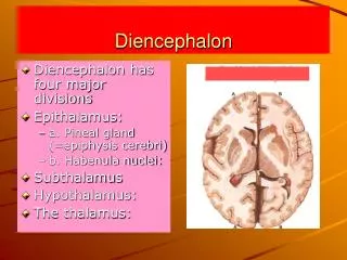
THE DIENCEPHALON • EPITHALAMUS • THALAMUS • SUBTHALAMUS • HYPOTHALAMUS
1- epithalamus 2 – thalamus 3- subthalamus 4 - hypothalamus
Diencephalon- Superior aspect
THE THALAMUS • Anterior nuclei • Medial nuclei (mediodorsal nc.) • Lateral nuclei – dorsal tier (lateral dorsal nc., lateral posterior nc. ventral tier ( ventral anterior – VA, ventral lateral – VL, ventral posterolateral- VPL, ventral posteromedial – VPM, ventral intermediate - VIM, • Medial geniculate nc., • Lateral geniculate nc., • Intralaminar nuclei • Midline nuclei • Posterior nuclei (nuclei of pulvinar) • Reticular nucleus
Thalamus – pulvinar and epithalamic nuclei (medial and lateral geniculate body)
Pul = pulvinar thalami MGB = medial geniculate body LGB = lateral ge niculate body
Thalamocortical and Corticothalamic loop input
Thalamic nuclei • Relay nuclei (specific) – MGN, LGN, VPL, VPM, VL, VA • Receives input predominantly from a single source • Processed information is sent to a localized region of cortex • Are modality specific • Specific nuclei (after stimulation sharply localized cortical response)
Association (nonspecific) nuclei • MD, LD, LP, Posterior ncc., • Receives input from a number of structures or cortical areas • Sends fibers to the association cortical areas
Auditory pathway + BG visual pathway trigemino-thalamictr. spino-thalamictr. lemniscalsystem Afferent connections of the main thalamic nuclei
Cerebello (dentato)-thalamic projections Majority of cerebellar fibers terminate in the VL nucleus Cerebello
No discrete nuclei Regulation of food and water intake Tuberal region VM – satiety center (lesion produces hyperphagia + obesity) Arcuate nc. - delivers peptides to the portal vessels Mamillary region Posterior nc.- elevating of blood pressure, pupillary dilatation, body heat conservation Mammillary ncc. – memory formation THE HYPOTHALAMUS • Lateral zone • Medial zone • Well defined nuclei • Chiasmatic region (hormone release- • SO,PV) • Cardiovascular function (Ant.) • Circadian rhytms (SCH) • Body temperature (Preoptic nc.)
References: P.Brodal : Central nervous system, Oxford, 2006 J. H. Martin : Neuroanatomy, McGraw-Hill, 2003 V. Chan - Palay : Cerebellar dentate nucleus, Springer 1977. P. Kopf – Meier. Atlas of Human Anatomy Karger, 2000.