Download
1 / 45
460 likes | 643 Views
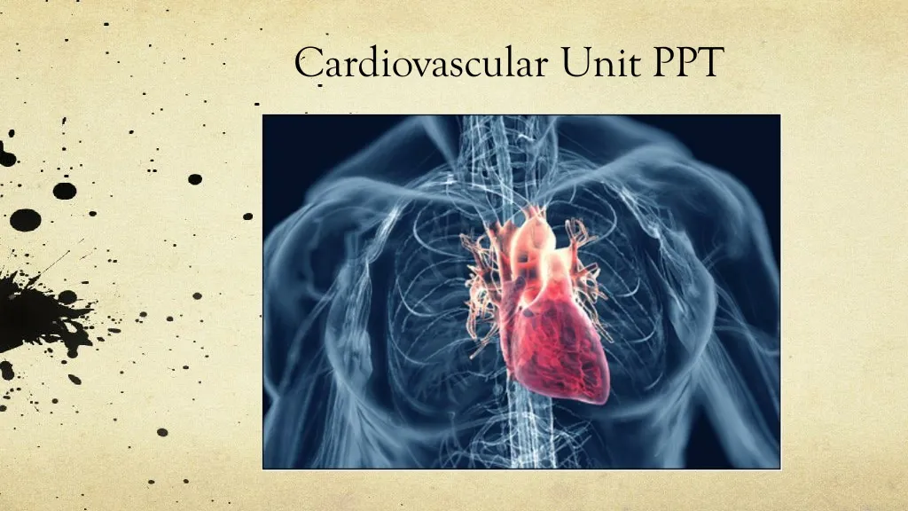
Cardiovascular Unit PPT. Make a Heart. You will need a worksheet Share instructions Colored pencils/markers Needs to be done by end of hour for use in next class. Circulatory Worksheet ( label #4-7 together). REMEMBER the difference between Veins and Arteries!!.

E N D
Make a Heart • You will need a worksheet • Share instructions • Colored pencils/markers • Needs to be done by end of hour for use in next class
Circulatory Worksheet F/U: • Looking at # 10 &11: The ventricles push blood out of the heart. If they are not working perfectly : • What is that persons body not receiving • What would the long term affects be • How could you treat it • On the back of Worksheet, list and define the directional terms • On the heart diagram label the anatomical structures that are on the worksheet. There should be 7+ (actually label the heart diagram)
Heart Anatomy: Flashcards: • You will need to: • Cut • Use BLUE, RED and BLACK colors
Cardiovascular Medical Abbreviations • Using Appendix B fill in chart • Please put packet back when finished
ADL Activities of Daily Living meds medication am Morning MI Myocardial infarction BLS Basic life support NKA No Known Allergies bpm Beats per minute NPO Nothing by mouth B/P, BP Blood pressure OR Operating room CCU Cardiac care unit preop Before surgery CHD Congenital heart defects, coronary heart disease postop After surgery CHF Congestive heart failure RR Respiratory rate CXR Chest x-ray stat immediately DOB Date of birth TIA Transient ischemic attack Dx diagnosis TV Tidal volume, tricuspid valve ECG/ EKG electrocardiogram unc. Unconscious Etiol etiologyvit vitamin inj injection w/c wheelchair
Medical abbreviations practice • Take a family history, date of birth, weight before examination. ______________________________________________________ 2. Record all vital signs, blood pressure, temperature and pulse three times a day ______________________________________________________ 3. Take chest xray, electrocardiogram before surgery ______________________________________________________ 4. Move patient to recovery room with wheelchair and give them bathroom privileges. ______________________________________________________ Take FH, DOB, wt before exam Record VS, BP T, P tid Take CXR, ECG/EKG preop Move pt RR c w/c BRP
Word Parts • Fill in what you already know from other units! • Use Appendix A for new word parts
Erythro- redArter/o artery Leuk- whiteAther/o plaque; fatty substance Tachy- fastAtri/o atrium cyte cellCardi/o, card/o heart -ary, -ic, ac pertaining to Valv/o valve Hem/o, hemat/o blood Phleb/o vein -itis inflammation of -lysis break down -malacia abnormal softening -osis abnormal condition of; disease -sclerosis abnormal hardening -stenosis abnormal narrowing Diastol/o expansion (relaxation) Systol/o contraction -verse, -version turn-ion action; process; state; or condition Hepat/o liverCirculat/o circulate
Circulatory Vocabulary • Using your Word Part chart fill in the vocab terms that you are able to • Make a GUESS and do your BEST!
Add to your Vocabulary: • Atherosclerosis: narrowing/hardening of blood vessels caused by deposits of fatty material containing calcium and cholesterol • Coronary: pertaining to the heart • Diastolic: dilation of the heart, resting phase or filling of the ventricles • Hepatic circulation: path of blood from the intestines, GB, pancreas, stomach and spleen THROUGH the liver • Pulmonary circulation: heart to lung. Carries de-oxygenated blood from the right ventricle to the lungs and returns oxygenated blood to the left atrium of the heart • Systemic circulation: general circ to systems. Oxygenated blood from the left ventricle to tissues of the body, returns de-oxygenated blood to right atrium • Systolic: contraction of the ventricles
Path of Blood = Blood Flow 3. Right atrium 4. Tricuspid valve 5. Right ventricle 2. superior/inferior vena cava 6. Pulmonary arteries 1. All parts of the body 7. lungs 12. aorta 8.Pulmonary veins 11. Left ventricle 10. bicuspid/mitral valve 9. Left atrium
Heart Circulation THREE TYPES • Pulmonary Circulation: Flow of blood between the heart and lungs • Systemic Circulation: Flow of blood between the heart and the cells of the body • Coronary Circuloation: Flow of blood within the heart
Blood Flow BLOOD VESSELS • Arteries carry blood AWAY from the heart - Largest = Aorta - Heart muscle contractions pump blood through arteries • Veins carry blood TOWARDS the heart - Largest = Superior/Inferior Vena Cava - Valves prevent blood from returning to heart - Skeletal muscle contractions move blood through veins
Blood Flow Continued… HEART VALVES • Controls blood flow • Valve between Left Atrium and Left Ventricle = bicuspid • Valve between Right Atrium and Right Ventricle = tricuspid • Pulmonary and Aortic Valves STOP the back flow of blood into the heart
Structures • HEART • Beats 72 times a MINUTE • 100,000 times a DAY • 3 Trillion times in a LIFETIME! • Circulates about 5-7 liters of blood • BLOOD VESSELS • Arteries • Veins
Functions • Transport nutrients and oxygen • Transport waste to kidneys • Distribute hormones and antibodies • Help control body temperature and maintain homeostasis
THE HEART • 2 Sided double pump • Is about the size of your fist • Lies in the Thoracic Cavity between the lungs
Heart Tissue • Endocardium: smooth membranous lining inside the heart • Myocardium: muscle tissue that is contractile, thickest layer
Heart Tissue Continued… • Epicardium: Outermost layer of the heart • Pericardium: covers the whole outside of the heart
Parts of the Heart • Divided into right and left sides • 2 chambers in each side, for a total of 4 chambers • Atrium: top, where blood enters (“top of tree”) • Ventricles: bottom, where blood leaves (“vents at the bottom”) • Left and right sides separated by a partition called a septum
Cardiac Conduction System • Electrical Impulses produce a wave that can be recorded on the ECG • Consists of… • Sinoatrial (SA) node • Atrioventriclular (AV) node • Bundle of His (AV Bundle) • Bundle Branches • Purkinje Fibers (network)
SA NODE • Located in the upper right part of the atrium • Is a natural pacemaker • Fires at a rate of 60 to 100 times per minute • The heartbeat starts in the SA node
AV NODE • Located in the floor of the right atrium • Delays or slows the electrical impulse • Fires at a rate of 40 to 60 time per minute • Can take over if the SA node is not working
Bundle of His • Located next to the AV node • Transfers the electrical impulse from the atria to the ventricles
Bundle Branches • Located along the left and right side of the intraventricular septum • Act as pathways or a fork in the road • Impulses in the bundle branch perform the important work of making the heart muscle contract
Purkinje Network • Provide an electrical pathway for each of the cardiac cells • Activate the left and right ventricles simultaneously causing the ventricles to contract
Pulse • Use Reading Packet to fill in the Pulse worksheet
Heart Sounds • Lubb Sound • Heard first • Mitral and tricuspid valves closing between the atria and ventricles • Dupp Sound • Heard second • Shorter and higher pitched • Closing of the aortic and pulmonary valves as blood is pumped out of the heart • Murmurs • Abnormal or extra sounds http://depts.washington.edu/physdx/heart/demo.html
Blood Pressure • Systolic = contraction of the ventricles • Diastolic = relaxation of the ventricles • Normal BP= 120/80 (systolic/diastolic) • Healthy systolic is less than 140 and greater than 90 • Healthy diastolic should be less than 100 http://www.nejm.org/doi/full/10.1056/NEJMvcm0800157
Blood Pressure Readings • Use Reading Packet fill in the Blood Pressure worksheet
Health Concerns/Assessments/Risk Factors • Using Teacher website • Click cardiovascular unit • Click website • Fill in guided notes using the website
Path of Blood: blood flow 3. 4. Tricuspid valve 5. 2. 6. 1. All parts of the body 7. 12. 8.Pulmonary veins 11. Left ventricle 10. 9.
There are _____ chambers of the heart • There are _____ tissue layers to the heart • The heart beats ____ times per minute • Average systolic BP _____ • Average diastolic BP ____ • Systolic BP range ____ • Diastolic BP range _____ • Average pulse _____