Download
1 / 16
210 likes | 1.78k Views
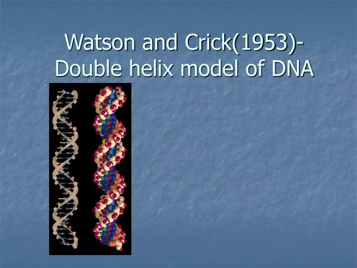
Watson and Crick(1953)- Double helix model of DNA. Double helix model of DNA. Early models of DNA.

E N D
Early models of DNA A structure for nucleic acid has already been proposed by Pauling and Corey. Their model consists of three intertwined chains, with the phosphates near the fibre axis, and the bases on the outside. Another three-chain structure has also been suggested by Fraser. In his model the phosphates are on the outside and the bases on the inside, linked together by hydrogen bonds.
On Feb. 28, 1953, Francis Crick walked into the Eagle pub in Cambridge, England, and, as James Watson later recalled, announced that "we had found the secret of life." Actually, they had. That morning, Watson and Crick had figured out the structure of deoxyribonucleic acid, DNA. And that structure — a "double helix" that can "unzip" to make copies of itself — confirmed suspicions that DNA carries life's hereditary information.
Francis Crick and James Watson with Maurice Wilkins received the 1962 Nobel Prize for discovering the molecular structure of deoxyribonucleic acid (DNA). • Widely regarded as one of the most important discoveries of the 20th century it has led the way to the mapping and deciphering of all the genes in the human chromosomes
X-ray diffraction • X-ray crystallography was originally used to look at the structures of simple organic minerals, but was progressively applied to more and more complex molecules. It aided in determining the structures of the alpha helix, the beta sheet, hemoglobin, and DNA
X-Ray Diffraction and the Structure of DNA • In 1951,, James Watson, joined the lab and the two formed a close working relationship. They were convinced that if the three-dimensional structure of a molecule known to play a role in passing genetic information -- DNA -- could be determined,. They made models based on research done in several fields. Crick and Watson saw the result of Rosalind Franklin's x-ray diffraction studies, and a final piece of the puzzle was fitted. In 1953 they created a visual model of DNA
X-Ray Diffraction and the Structure of DNA • They made models based on research done in several fields. Crick and Watson saw the result of Rosalind Franklin's x-ray diffraction studies, and a final piece of the puzzle was fitted. In 1953 they created a visual model of DNA
X-Ray Diffraction and the Structure of DNA Watson was shown this picture by Wilkins in early 1953. From the picture it was possible to calculate: 1) the distance between bases (3.4A) 2) the length of the period (34A) 3) the rise of the helix (36 degrees)
Building model • Therefore, knowing that DNA existed and contained four bases, a ribose sugar and phosphate. Inspired by Pauling's successful attempts at building 3-D models of proteins, Crick and Watson believed this to be the correct way to proceed.
Bases • John Griffith, the mathematician nephew of Fred Griffith, calculated the attractive forces between 'like' bases. Crick's idea was that since the bases were flat, perhaps they coud be stacked on top of one another, and attracted that way.Griffith informed him that adenine attracted thymine and guanine attracts cytosine
Chargaff’s 1:1 Rule A:T=G:C+1:1 When Crick compared the bases that Griffiths had told him with Chargraff's data, he realized that complementary base pairing could be the cause of the 1:1 rule.
Hydrogen bond • Watson and Crick was the thought that hydrogen bonding was too unstable to be responsible for replication. Crick was also assuming that both tautameric forms of the bases existed in the same DNA molecule, and that the proton could shift from one position to another, thus altering the sites for hydrogen bond formation.
The final model • This structure has two helical chains each coiled roundthe same axis • usual chemical assumptions, namely, that each chain consists of phosphate diester groups joining ß-D-deoxyribofuranose residues with 3',5' linkages • Both chains follow right- handed helices, but owing to the dyad the sequences of the atoms in the two chains run in opposite directions
The final model • an angle of 36 degrees between adjacent residues in the same chain, so that the structure repeats after 10 residues on each chain, that is, after 34 A. The distance of a phosphorus atom from the fibre axis is 10 A. • As the phosphates are on the outside, cations have easy access to them • The phosphates are negatively charged, and attract cations. The phosphates, being charged, are also hydrophilic
For their outstanding work in discovering the double helical structure of DNA, Watson and Crick shared the 1962 Nobel Prize for Physiology and Medicine with Maurice Wilkins. Sadly, Rosalind Franklin, whose work greatly contributed to this key discovery