Download
1 / 3
30 likes | 32 Views
MRI: Magnetic Resonance Imaging; CT-scan: Computed Tomography Scan; LE: Limbic Encephalitis; MoCA: Montreal Cognitive Assessment; PNS: Paraneoplastic Neurological Syndromes; CSF: Cerebro-Spinal Fluid; GABA: Gamma-Aminobutyric Acid Receptor; VGCC: Voltage-Gated Calcium Channels; VGKC: Voltage-Gated Potassium Channels

E N D
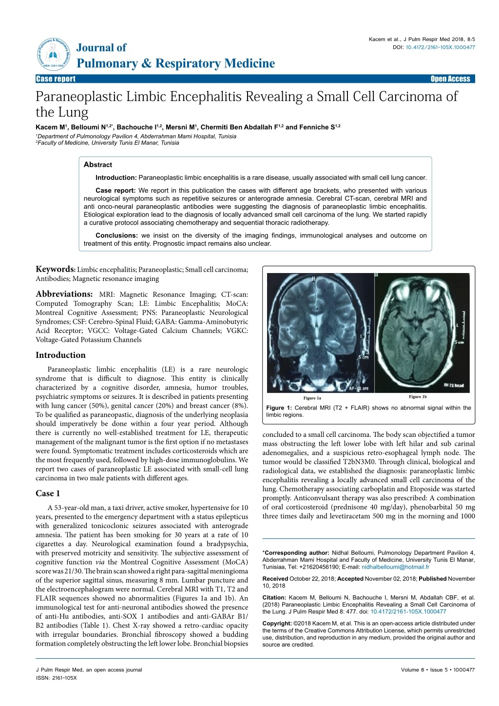
Kacem et al., J Pulm Respir Med 2018, 8:5 DOI: 10.4172/2161-105X.1000477 JournalofPulmonary&RespiratoryMedicine ISSN: 2161-105X Journal of Pulmonary & Respiratory Medicine Case report Paraneoplastic Limbic Encephalitis Revealing a Small Cell Carcinoma of the Lung Kacem M1, Belloumi N1,2*, Bachouche I1,2, Mersni M1, Chermiti Ben Abdallah F1,2 and Fenniche S1,2 Open Access 1Department of Pulmonology Pavilion 4, Abderrahman Mami Hospital, Tunisia 2Faculty of Medicine, University Tunis El Manar, Tunisia Abstract Introduction: Paraneoplastic limbic encephalitis is a rare disease, usually associated with small cell lung cancer. Case report: We report in this publication the cases with different age brackets, who presented with various neurological symptoms such as repetitive seizures or anterograde amnesia. Cerebral CT-scan, cerebral MRI and anti onco-neural paraneoplastic antibodies were suggesting the diagnosis of paraneoplastic limbic encephalitis. Etiological exploration lead to the diagnosis of locally advanced small cell carcinoma of the lung. We started rapidly a curative protocol associating chemotherapy and sequential thoracic radiotherapy. Conclusions: we insist on the diversity of the imaging findings, immunological analyses and outcome on treatment of this entity. Prognostic impact remains also unclear. Keywords: Limbic encephalitis; Paraneoplastic; Small cell carcinoma; Antibodies; Magnetic resonance imaging Abbreviations:MRI: Magnetic Resonance Imaging; CT-scan: Computed Tomography Scan; LE: Limbic Encephalitis; MoCA: Montreal Cognitive Assessment; PNS: Paraneoplastic Neurological Syndromes; CSF: Cerebro-Spinal Fluid; GABA: Gamma-Aminobutyric Acid Receptor; VGCC: Voltage-Gated Calcium Channels; VGKC: Voltage-Gated Potassium Channels Introduction Paraneoplastic limbic encephalitis (LE) is a rare neurologic syndrome that is difficult to diagnose. This entity is clinically characterized by a cognitive disorder, amnesia, humor troubles, psychiatric symptoms or seizures. It is described in patients presenting with lung cancer (50%), genital cancer (20%) and breast cancer (8%). To be qualified as paraneopastic, diagnosis of the underlying neoplasia should imperatively be done within a four year period. Although there is currently no well-established treatment for LE, therapeutic management of the malignant tumor is the first option if no metastases were found. Symptomatic treatment includes corticosteroids which are the most frequently used, followed by high-dose immunoglobulins. We report two cases of paraneoplastic LE associated with small-cell lung carcinoma in two male patients with different ages. Case 1 A 53-year-old man, a taxi driver, active smoker, hypertensive for 10 years, presented to the emergency department with a status epilepticus with generalized tonicoclonic seizures associated with anterograde amnesia. The patient has been smoking for 30 years at a rate of 10 cigarettes a day. Neurological examination found a bradypsychia, with preserved motricity and sensitivity. The subjective assessment of cognitive function via the Montreal Cognitive Assessment (MoCA) score was 21/30. The brain scan showed a right para-sagittal meningioma of the superior sagittal sinus, measuring 8 mm. Lumbar puncture and the electroencephalogram were normal. Cerebral MRI with T1, T2 and FLAIR sequences showed no abnormalities (Figures 1a and 1b). An immunological test for anti-neuronal antibodies showed the presence of anti-Hu antibodies, anti-SOX 1 antibodies and anti-GABAr B1/ B2 antibodies (Table 1). Chest X-ray showed a retro-cardiac opacity with irregular boundaries. Bronchial fibroscopy showed a budding formation completely obstructing the left lower lobe. Bronchial biopsies Figure 1: Cerebral MRI (T2 + FLAIR) shows no abnormal signal within the limbic regions. concluded to a small cell carcinoma. The body scan objectified a tumor mass obstructing the left lower lobe with left hilar and sub carinal adenomegalies, and a suspicious retro-esophageal lymph node. The tumor would be classified T2bN3M0. Through clinical, biological and radiological data, we established the diagnosis: paraneoplastic limbic encephalitis revealing a locally advanced small cell carcinoma of the lung. Chemotherapy associating carboplatin and Etoposide was started promptly. Anticonvulsant therapy was also prescribed: A combination of oral corticosteroid (prednisone 40 mg/day), phenobarbital 50 mg three times daily and levetiracetam 500 mg in the morning and 1000 *Corresponding author: Nidhal Belloumi, Pulmonology Department Pavilion 4, Abderrahman Mami Hospital and Faculty of Medicine, University Tunis El Manar, Tunisiaa, Tel: +21620456190; E-mail: nidhalbelloumi@hotmail.fr Received October 22, 2018; Accepted November 02, 2018; Published November 10, 2018 Citation: Kacem M, Belloumi N, Bachouche I, Mersni M, Abdallah CBF, et al. (2018) Paraneoplastic Limbic Encephalitis Revealing a Small Cell Carcinoma of the Lung. J Pulm Respir Med 8: 477. doi: 10.4172/2161-105X.1000477 Copyright: ©2018 Kacem M, et al. This is an open-access article distributed under the terms of the Creative Commons Attribution License, which permits unrestricted use, distribution, and reproduction in any medium, provided the original author and source are credited. J Pulm Respir Med, an open access journal ISSN: 2161-105X Volume 8 • Issue 5 • 1000477
Citation: Kacem M, Belloumi N, Bachouche I, Mersni M, Abdallah CBF, et al. (2018) Paraneoplastic Limbic Encephalitis Revealing a Small Cell Carcinoma of the Lung. J Pulm Respir Med 8: 477. doi: 10.4172/2161-105X.1000477 Page 2 of 3 Antibodies Results Antibodies Results Anti Cv2 - Anti Titin - Anti PNMA - Anti Amphiphysin - Anti Ri - Anti AMPA1/AMPA2 - Anti Yo - Anti CASPR2 - Anti Hu + Anti LG11 - Anti Recoverin - Anti GABAr B1/B2 ++ Anti SOX1 ++ Table 1: Serum immunoassay for anti-onco-neuronal and anti- membrane antibodies. mg at night. Despite the treatment, the patient had a seizure every two weeks. The onset of chemotherapy had a positive impact with disappearance of the seizures. During chemotherapy sessions, the patient was still bradypsychic but with a more sustained memory. The MoCA score was 25/30. After 4 cycles of chemotherapy based on carboplatin and etoposide, we noted a stability of the tumor. Therefore, sequential thoracic radiotherapy was proposed. Case 2 A 73 years old man, former smoker, was admitted to pulmonology department for exploration of a chronic dry cough. The patient has been smoking for 42 years at a rate of 10 to 20 cigarettes a day. He had past medical history of a treated gastric ulcer. He was complaining of progressively emerging cough with retrosternal burn sensation. His family members signaled anterograde amnesia with neither humor trouble nor suicidal tendency. Physical examination showed a normal cardio-pulmonary status, normal sensitivity and motricity. Chest X-ray revealed a right hilar opacity with spiculated margins. Bronchial fibroscopy showed a budding formation partially obstructing the right upper lobar bronchus. Bronchial biopsies concluded to small cell carcinoma. The thoracic CT scan objectified a tissue mass extending from the hilus to the right upper lobe, measuring 59 mm of diameter, associated with sub-pleural speculated nodules of the right lower lobe, mediastinal lymph nodes in the zones 4R, 7 and 10 (Figure 2). Cerebral MRI revealed bilateral high signal intensity on T2-weighted and FLAIR image in the hippocampus (Figures 3a and 3b). We concluded on paraneoplastic limbic encephalitis. The tumor was classified T4N3M0 according to the 8th edition of TNM classification. No treatment was prescribed for the encephalitis. Four cycles of chemotherapy including carboplatin and etoposide, then sequential thoracic radiotherapy was proposed. Discussion Paraneoplastic neurological syndromes (PNS) are rare neurological syndromes associated with cancer that are not explained by a metastatic, metabolic, infectious, deficiency or iatrogenic cause [1]. The pathogenesis of this condition is not completely understood, but the most likely hypothesis is that of an immune response directed against the antigens expressed by the nervous system cells similar to the tumor antigens, resulting in neuronal loss with lymphocyte infiltration of perivascular and microglial cells [1]. The presence of circulating serum autoantibodies or abnormal cerebrospinal fluid (CSF), specifically associated with PNS is one of the hallmarks of these syndromes. They are found in more than 80% of patients with a PNS. Depending on the target of the antibodies found, there are two types of PNS. There are intracellular targets (onco-neuronal) and membrane targets [1]. Well- characterized anti-onco-neuronal antibodies such as anti-Hu, anti- Ri, anti-Yo, anti-Ma/Ta, anti-amphiphysin, anti-Sox1 and anti-CV-2 are true markers of cancer, generally small cell lung cancer, breast, ovarian or testicular cancer. Clinical data and the specificity of the anti- Figure 2: Thoracic CT shows a tumor mass that obstructs the left lower lobe bronchus with left hilar and sub-carinal adenomegalies. Figure 3: Cerebral MRI revealing bilateral high signal intensity on T2-weighted and FLAIR image in the hippocampus. onco-neuronal antibodies guide the oncological assessment [2]. The best described membrane targets are ionotropic glutamate receptors: N-Methyl D-Aspartate Receptor (NMDAr) or Gamma-Aminobutyric Acid Receptor (GABAr), either ion channels such as voltage-gated calcium channels (VGCC) or voltage-gated potassium channels (VGKC) [2]. The clinical presentations vary from case to case. The onset is mostly sub-acute. In 65% of cases, paraneoplastic neurological involvement precedes the discovery of cancer for several months or years. Autoimmune encephalitis is one of the most common PNS, involving mainly the limbic system but may also involve other extra limbic structures. The term autoimmune encephalitis is preferred to LE which is too restrictive [1]. Neurological symptoms vary from anterograde memory disorders, confusion to dementia and convulsive seizures that may precede the cognitive impairment for several months (temporal, psychomotor impairment, generalized tonic-clonic seizures). Changes of behavior, personality and psychiatric symptoms (irritability, anxiety, depression, hallucinations and aboulia) are also described. Sleep disorders (hypersomnia or insomnia, narcolepsy, cataplexy), weight change along with disturbance of the sensation of satiety can be found [3-5]. Further tests are necessary to make the diagnosis but also to rule out the differential diagnoses (infectious cause, deficiency, metastatic). The analysis of the cerebrospinal fluid contributes to the diagnosis by showing the absence of malignant cells. J Pulm Respir Med, an open access journal ISSN: 2161-105X Volume 8 • Issue 5 • 1000477
Citation: Kacem M, Belloumi N, Bachouche I, Mersni M, Abdallah CBF, et al. (2018) Paraneoplastic Limbic Encephalitis Revealing a Small Cell Carcinoma of the Lung. J Pulm Respir Med 8: 477. doi: 10.4172/2161-105X.1000477 Page 3 of 3 Cerebral MRI shows no metastatic lesion. The electroencephalogram shows a non-specific aspect of epileptic activities at the temporal lobes in 50% of cases [6]. MRI plays also a prominent role in diagnosis by detecting single or bilateral (60%) amygdalo-hippocampal signal abnormalities [3,6]. Typically, imaging shows a high-intensity signal on T2-weighting sequences, more visible on the FLAIR and on the diffusion sequences. T1-weighting sequences may show a hyposignal or isosignal with temporal atrophy at a later stage. Enhancement after gadolinium injection can be seen in 15 to 20% of cases [3,6]. MRI may be normal at the beginning, as is the first case, hence the importance of imaging follow-up [7]. FDG-PET scan is useful when the EEG and MRI show no abnormalities. It shows a hyper-metabolism of the temporal lobes, midbrain, cerebellum or frontotemporal lobes [8], indicating an acute phase of the inflammatory process. The presence of anti-neuronal anti-GABAr B1 antibodies is suggestive of small cell cancer. The presence of anti-Hu antibodies is associated in 94% of cases with small- cell lung carcinoma [3]. Alamowitch et al. [9] found anti-Hu antibodies in 50% of patients with limbic encephalitis associated with small cell lung carcinoma. Anti-SOX1 antibodies are found in the Lambert- Eaton myasthenic syndrome and less frequently in patients with anti- Hu syndrome or small-cell lung carcinoma without neurological signs [10]. GABAr B antibody assay should be considered in all patients with LE with or without associated small cell carcinoma, and in cases of cerebellar dysfunction and opsoclonus-myoclonus without other identified antibodies. The evolution and prognosis of paraneoplastic LE depends on the nature of the primary tumor and its treatment. The presence of anti-neuronal antibodies is often associated with poor neurological functional prognosis despite the proposed therapeutic arsenal. It is currently considered that the treatment of the tumor is the only one able to stabilize the evolution by suppressing the antigenic stimulation [1]. In the presence of a membrane antibody, treatments targeting humoral immunity (immunoglobulin IV, plasmapheresis, and anti-CD20) can be proposed as well as systemic corticosteroids. However, no therapeutic protocol could be validated by a randomized controlled study because of the small number of patients concerned [1]. Conclusion The reported cases highlight the diversity of paraneoplastic syndromes, which can be the first symptom of a malignant tumor. The review of the literature confirms the crucial importance of the anti-tumor immune response that can generate temporary or permanent dysimmunitary or endocrine disorders. The complexity of the pathophysiology explains the difficulty of treatment. It is also difficult to describe predictive elements of response of paraneoplastic neurological signs to the proposed therapies, apart from regression of the tumor. Tumor control remains the best alternative. Author’s Contributions Kacem M: 1st case redaction, scientific content discussion Belloumi N: Article redaction, correction, scientific content discussion Bachouche I: Scientific content discussion Mersni M: 2nd case redaction Chermiti Ben Abdallah F: Scientific content discussion Fenniche S: Scientific content discussion Consent for Publication A written consent for publication was obtained from the patients and their legal tutors. Competing Interests No competing interests related to the manuscript theme. References 1. Joubert B, Honnorat J (2014) Syndromes neurologiques paranéoplasiques. La Lettre du Neurologue 3: 96-101. 2. Goetz J, Olsson NO, Humbel RL (2013) Anticorps antineuronaux. EMC Biologie médicale. 3. Gultekin SH, Rosenfeld MR, Voltz R, Eichen J, Posner JB (1000) Paraneoplastic limbic encephalitis: Neurological symptoms, immunological findings and tumour association in 50 patients. Brain 123: 1481-1494. 4. Honnorat J, Antoine JC (2007) Paraneoplastic neurological syndromes. Orphanet J Rare Dis 2: 22. 5. Dalmau J, Bataller J (2006) Clinical and immunological diversity of limbic encephalitis: A model for paraneoplastic neurologic disorders. Hematol Oncol Clin North Am 20:1319-1335. 6. Lawn ND, Westmoreland BF, Kiely MJ, Lennon VA, Vernino S (2003) Clinical, magnetic resonance imaging, and electroencephalographic findings in paraneoplastic limbic encephalitis. Mayo Clin Proc 78: 1363-1368. 7. Messori A, Lanza C, Serio A, Salvolini U (2003) Resolution of limbic encephalitis with detection and treatment of lung cancer: Clinical-radiological correlation. Eur J Radiol 45: 78-80. 8. Ances BM, Vitaliani R, Taylor RA, Liebeskind DS, Voloschin A, et al. (2005) Treatment-responsive limbic encephalitis identified by neuropil antibodies: MRI and PET correlates. Brain 128: 1764-1777. 9. Alamowitch S, Graus F, Uchuya M, René R, Bescansa E, et al. (1997) Limbic encephalitis and small cell lung cancer, clinical and immunological features. Brain 120: 923-928. 10. Tschernatsch M, Singh P, Gross O, Gerriets T, Kneifel N, et al. (2010) Anti- SOX1 antibodies in patients with paraneoplastic and non-paraneoplastic neuropathy. J Neuroimmunol 226: 177-180. J Pulm Respir Med, an open access journal ISSN: 2161-105X Volume 8 • Issue 5 • 1000477