Download
1 / 60
631 likes | 749 Views
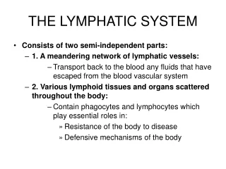
the lymphatic system. consists of The lymph conducting channels Lymphoid tissues Lymphoid organs. The lymphatic system is part of the immune system and acts as a secondary (accessory) circulatory system. Functions of the lymphatic system:. remove excess fluids from body tissues,

E N D
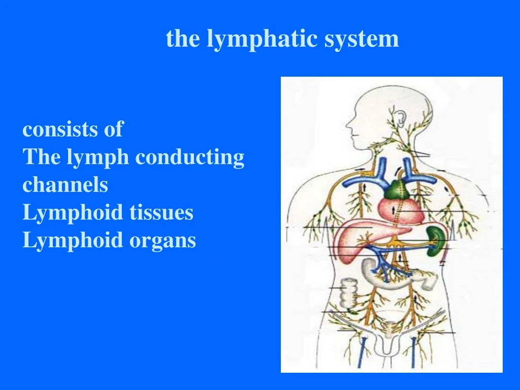
the lymphatic system consists of The lymph conducting channels Lymphoid tissues Lymphoid organs
The lymphatic system is part of the immune system and acts as a secondary (accessory) circulatory system.
Functions of the lymphatic system: • remove excess fluids from body tissues, • absorb fatty acid and transport fat to circulatory system, and • produce immune cells (lymphocytes, monocytes, and plasma cells).
Blood fluid escapes through the thin-walled capillaries into spaces between body tissue cells. Lymph vessels, which have very thin walls, pick up these fluids called lymph.
Composition • Lymphatic vessel • Lymphatic capillary • Lymphatic vessels: two sets, superficial and deep • Lymphatic trunks: nine • Lymphatic ducts: thoracic duct, right lymphatic duct • Lymphatic organ • Lymphatic nodes • Tonsil, spleen, thymus • Lymphatic tissue • Include diffused lymphoid tissues and lymph nodules. They are mainly situated in the wall of the respiratory-alimentary tracts and consist of aggregated of lymphocytes and associates cells.
Tissue fluid and its formation • composition same as blood butwithout read blood cells (RBCs), platelets & proteins because they are too large to leak out of the capillaries - forms a link between blood and cells, providing a medium for exchange of materials between blood & cells
At the arterial endof a capillary, liquid is forced out as tissue fluid which is similar to plasma in composition except its has no plasma proteins, platelets & RBCs. At the venous end, some fluid returns to blood while some enters lymph vessels which eventually join to a vein near the heart and thus returns to blood finally.
Lymph some tissue fluid returns to capillaries by osmosis while some (about 10%) goes into lymph capillaries; this fluid is now called lymph Path: Blood lymph capillaries lymph vessels lymph ducts Blood - Lymph re-enters blood
Lymphatic capillaryBegin blindly • The wall is composed of a single layer of overlapping endothelial cells • They are numerous and form complex networks • The brain, spinal cord, bone marrow, parenchyma of spleen and eyeball lack lymphatic capillaries
Lymph is driven by contraction of surroundingmuscles, aided by valves which enable one-way flow to the neck.
Movement of lymph through the lymphatic system: 1. Hydrostatic pressure 2. Muscle contraction 3. Inspiratory movement 4.Valves to ensure one-way traffic towards the heart
The Lymphatic System Heart Vein Lymphatic duct Artery Lymphatic trunk Capillaries Lymphatic node Cell Tissue fluid Lymphatic capillary Lymphatic vessel
lymphatic vessels • lymphatic capillary • lymphatic vessel • lymphatic trunks • lumbar trunks(2) • brochomediastinal trunks(2) • subclavian trunks(2) • jugular trunks(2) • intestinal trunk • lymphatic ducts • thoracic duct • right lymphatic duct
Lymphatic vessel Features of structure • Three layered wall similar to, but thinner than vein, • More numerous valves than in vein • Interposed by lymph node at interval along the course • Arranged in superficial and deep sets
Lymphatic trunks • right and left jugular trunks • right and left subclaviantrunks • right and left bronchomediastinaltrunks • right and left lumbar trunks • intestinal trunk
Lymphatic ducts Right lymphatic duct • Formed by union of right jugular, subclavian, and bronchomediastinal trunks • Ends by entering the right venous angle • Receives lymph from right half of head, neck, thorax and right upper limb
Thoracic duct • Begins in front of L1 as a dilated sac, the cisterna chyli , which formed by joining of left and right lumbar trunks and intestinal trunk • Enter thoracic cavity by passing through the aortic hiatus of the diaphragm and ascends along on the front of the vertebral column, between thoracic aorta and azygos vein
Thoracic duct • Travels upward, veering to the left at the level of T5 • At the roof of the neck, it turns laterally and arches forwards and descends to enter the left venous angle • Just before termination, it receives the left jugular, subclavian and bronchomediastinal trunks
Thoracic duct • Drains lymph from lower limbs, pelvic cavity, abdominal cavity, left side of thorax, and left side of the head, neck and left upper limb
Lymphatic vessel • Have valves that give them a beaded appearance • Two sets: superficial (lie in the superficial fascia ) and deep (run with blood vessels and nerves) • Passes through at least one lymph node and often several
lymphatic ducts • thoracic duct from cisterna chyli (L1) to left venous angles • left jugular trunk • left subclavian trunk • left bronchomediastinal trunk • right and left lumbar trunk • intestinal trunk right lymphatic duct • right brochomediastinal trunk • right subclavian trunk • right jugular trunk
The lymph vessels join to form larger ducts that pass through lymph nodes (or glands). Each lymph node has a fibrous outer covering (capsule), a cortex, and a medulla.
lymph node (Small oval or bean-shaped bodes • Afferent vessels enter the node on its convex surface, and efferent vessels leave the node at its concave surface-the hilum • Arranged in groups, along the blood vessels • Regional nodes • is the lymph node where the lymph of the organ or part of the body drainage to firstly
Lymph nodes filter foreign substances, such as bacteria and cancer cells, from the lymph before it is re-entered into the blood system through the larger veins. Lymph nodes, which are scattered among the lymph vessels, act as the body’s first defense against infection.
Lymph nodes produce the following cells: • Lymphocytes – a type of white blood cell, • Monocytes – a leukocyte that protects against blood-borne pathogens, and • Plasma cells – produce antibodies.
Each lymph node has its own blood supply and venous drainage. The lymph nodes usually have names that are related to their location in the body.
When a specific location gets infected, the lymph nodes in that area will enlarge to fight the infection. If the lymph node closest to an infected area is unable to eliminate the infection, other lymph nodes in the system will attempt to fight the infection.
This is particularly critical in the case of cancer, which can be spread from its point of origin to all parts of the body through the lymphatic system.
The lymphatic drainageof head Lymph nodes of head • Located at junction of head and neck • Consist of • Occipital lymph nodes • Mastoid lymph nodes • Parotid lymph nodes ★Submandibular lymph nodeslies near the submandibular gland, receive lymphatic vessels from the face, nose and mouth • submental lymph nodes • Drain into deep cervical lymph nodes
Lymph nodes of the neck Anterior cervical ln. • Superficial anterior cervical lymph nodes • Deep anterior cervical lymph nodes Lateral cervical ln. ★Superficial lateral cervical ln. -lie along the external jugular vein ★ Deep lateral cervical ln. -extend along the internal jugular vein
Lymph nodes of the neck ★Deep lateral cervical ln. • Extend along the internal jugular vein from the base of skull to the root of neck • Divided into superior deep lateral cervical ln. and inferior deep lateral cervical ln. • Receive lymphatic vessels from head, neck, tongue, larynx, cervical parts of esophagus and trachea, thyroid gland, upper parts of the thoracic wall and breast • Efferent vessels form the jugular trunk • Left jugular trunk joins the thoracic duct • Right jugular trunk joins the right lymphatic duct
Lymph nodes of the neck Superior deep lateral cervical ln.Jugulodigastric ln. • Lies at the junction of posterior belly of digastric and internal jugular vein • Drain the nasopharynx, palatine tonsil and root of tongue Inferior deep lateral cervical ln.Juguloomohyoid ln. • Lies at the junction of the intermediate tendon of omohyoid and internal jugular vein • Drain the apex of tongue
Lymph nodes of the neck Inferior deep lateral cervical ln. Supraclavicular lymph nodes • Lie along transverse cervical a. & v. • palpable in the supraclavicular fossa. The most notable supraclavicular lymph node is Virchow's node. which can contain metastasis of visceral (abdominal) tumor. • Retropharyngeal ln. • Lying vertically behind the pharynx • drain nasopharyngeal lymph
Lymph nodes of upper limb • Cubital lymph node • lies above medial epicondyle of humerus • Receive lymph vessels from forearm • Axillary lymph node • arranged in five groups
Axillary lymph nodes • Axillary lymph nodes vary in size from a pin-head to a large bean. • They are arranged in five groups.
Axillary lymph nodes Pectoral lymph nodesLying along the lower border of pectoralis minor behind the pectoralis major • Receive lymph vessels from the lateral quadrants of the breast and superficial vessels from the anterolateral abdominal wall above the level of the umbilicus
Axillary lymph nodes Lateral lymph nodesAlong medial side distal part of axillary vein • Receives lymph from upper limb
Axillary lymph nodes Subscapular lymph node • Lying along subscapular vessels, in front of the subscapularis • Receive superficial lymph vessels from the back, down as far as the level of the iliac crests • Efferents of above three groups pass to central lymph node
Axillary lymph nodes Central lymph node • Lying in the center of the axilla in the axillary fat • Receive lymph from the above three nodes • Efferents pass to apical lymph node
Axillary lymph nodes Apical lymph node infraclavicular lymph nodes Lying at the apex of the axilla at the lateral border of the fist rib • Receive lymph of the efferent lymph vessels from all the other axillary nodes • The efferents of the apical nodes form the subclavian trunk
Axillary lymph nodes Efferents form subclavian trunk, the right subclavian trunk joins the right lymphatic duct; left usually drains directly into thoracic duct Apical ln. Subclavian trunk Central ln. Pectoral ln. Lateral ln. Subscapular ln.
Lymphatic drainage of thorax The lymphatic drainage of thoracic wall • To axillary lymph nodes • To parasternal lymph nodes (along internal thoracic vessels) • To intercostals lymph nodes from deeper structures
The lymphatic and venous drainages of the breast are of great importance in the spread of carcinoma. • About three quarters of the lymphatic drainage is to the axillary nodes: (1) Lymphatics pass around the edge of the pectoralis major and reach the pectoral group of axillary nodes; • (2) routes through or between the pectoral muscles may lead directly to the apical nodes of the axilla; • (3) lymphatics follow the blood vessels through the pectoralis major and enter the parasternal (internal thoracic) nodes; • (4) connections may lead across the median plane and hence to the contralateral breast; • (5) lymphatics may reach the sheath of the rectus abdominis and the subperitoneal and subhepatic plexuses. • It should be noted that free communication exists between nodes below and above the clavicle and between the axillary and cervical nodes.
lymph nodes of the thorax • Pulmonary ln. lie in the angles of bifurcation of branching lobar bronchi • Bronchopulmonary hilar ln.-lie in the hilums of the lung • Tracheobronchial ln.-situated above or below the bifurcation of trachea • Paratracheal ln.-along each side of the trachea
lymph nodes of the thorax • Anterior mediastinal lymph node • Lies anterior to the large blood vessels of thoracic cavity and pericardium • The efferents unite with those of paratracheal lymph nodes and parasternal lymph nodes to form the right and left bronchomediastinal trunks • The left bronchomediastinal trunk terminates in thoracic duct, and right in the right lymphtic duct • Posterior mediastinal lymph nodes lie along the esophagus and thoracic aorta
Lymph nodes of lower limb Popliteal ln. • Embedded in the fatty connective tissue of popliteal fossa • Receive superficial lymphatic vessels from posterolateral part of calf, and from deep lymphatic vessels accompanying anterior and posterior tibia a. • Efferents pass to the deep inguinal ln.
Lymph nodes of lower limb Superficial inguinal lymph nodesSuperior group: • Lies just distal to the inguinal ligament • Receive lymph vessels from anterior abdominal wall below umbilicus, gluteal region, perineal region, external genital organs • Inferior group: • Lies vertical along the terminal great saphenous v. • Receives all superficial lymph vessels of lower limb, except for those from the posterolateral part of calf • Efferent vessels drain into the deep inguinal ln. or external iliac ln.
Lymph nodes of lower limb Deep inguinal lymph nodes • Lie medial to the femoral v. • Receive deep lymph vessels of lower limb, perineal region, and efferent vessels from the superficial inguinal ln. • Drain into the external iliac ln.