Download
1 / 25
330 likes | 817 Views
TB, Lung Abscess, and Cystic Fibrosis. TB. Radiographic findings in primary TB are Nonspecific Tends to like the lower lung zones Cavitation is not as common in primary TB as in reactivation TB However lymphadenopathy is a common finding in primary TB and uncommon in reactivation TB.

E N D
TB • Radiographic findings in primary TB are Nonspecific • Tends to like the lower lung zones • Cavitation is not as common in primary TB as in reactivation TB • However lymphadenopathy is a common finding in primary TB and uncommon in reactivation TB
TB • Patchy left lower lobe opacity • Looks like pneumonia
TB • Right upper and lower lobe consolidation • Right pleural effusion
TB • Cavitary right upper lobe lesion • Right paratracheal lymphadenopathy • Right middle lobe infiltrate • Notice the ipsilateral lymphadenopathy
TB • Thick walled cavity with satellite nodules • Smooth inner wall
TB • Focal right middle lobe infiltrate • Nodular like infiltrate • Endobronchial spread of TB • Adjacent areas of lung are infected by bronchial secretions
TB • Radiographic findings usually present 2 years after initial infection • Infiltrates usually like the apical and posterior segments of upper lobes and superior segment of lower lobes
TB • CT scan through the upper chest shows a thick walled cavity with an air fluid level and surrounding infiltrate • Cavities result from caseous necrosis
TB • Complications of TB cavities • Mycetoma “fungus ball” • Rasmussen Aneurysm which is weakening of bronchial artery adjacent to a cavitary lesion
TB • Bilateral lung nodules resulting from endobronchial spread of TB • Right upper lobe cavity
Miliary TB • Right paratracheal lymphadenopathy • Bilateral tiny uniform nodules • Diffuse pattern of nodules is due to hematogenous spread
TB Key Points • Imaging findings of primary TB are nonspecific • Primary TB differentiated from bacterial pneumonia by the presence of lymphadenopathy • Reactivation TB recognized by fibrocavitary disease and a history of prior exposure
TB Key Points • Inactive disease cannot be established without prior films • Primary TB tends to affect the lower lungzones while reactivation TB tends to affect the upper lung zones
Pneumococcal PNA • Complications • Lung necrosis • Abscess formation • Often need clinical history to distinguish from TB
Lung Abscess • Air fluid level within a large cavity • Can communicate with the pleura resulting in an empyema
Lung Abscess • 54 year old male with cough and foul smelling sputum • Cavity within the superior segment of the left lower lobe • Common site for aspiration
Lung Abscess • Irregular cavity • Typically more posterior • Often has an air/fluid level within it • Often has surrounding infiltrate
Lung Abscess • Cavity with air fluid level and foul smelling sputum • Anaerobic organisms often the cause of abscesses from aspiration
Lung Abscess Key Points • Typical radiographic appearance is an irregular cavity with an air fluid level • Lung abscesses from aspiration often occur in the posterior segments of upper lobes or superior segments of lower lobes • The wall thickness of lung abscesses progresses from thick to thin and irregular to well circumscribed
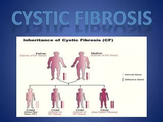
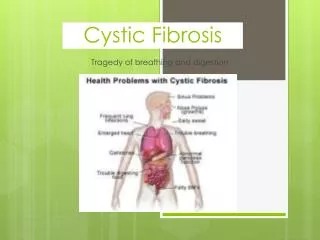
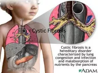
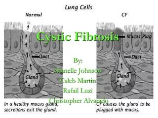
Cystic Fibrosis • Abnormal sodium/chloride transport in exocrine tissues • Results in thick viscous mucus • Obstructs airways resulting in repeat infections and colonization • Airways dilate and cysts form from air trapping • Scarring from the repeated infections
Cystic Fibrosis • Hyperinflation • Upper lobe bronchiectasis • Tram tracking