Download
1 / 83
830 likes | 902 Views
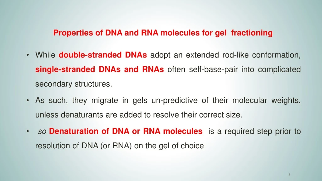
Properties of DNA and RNA molecules for gel f ractioning. While double-stranded DNAs adopt an extended rod-like conformation, single-stranded DNAs and RNAs often self-base-pair into complicated secondary structures.

E N D
Properties of DNA and RNA molecules for gel fractioning • While double-stranded DNAs adopt an extended rod-like conformation, single-stranded DNAs and RNAs often self-base-pair into complicated secondary structures. • As such, they migrate in gels un-predictive of their molecular weights, unless denaturants are added to resolve their correct size. • so Denaturation of DNA or RNA moleculesis a required step prior to resolution of DNA (or RNA) on the gel of choice
Secondary RNA and DNA Structures RNA Secondary Structures tRNA Structure Credit: David Marcey Cal Lutheran Univ.
CELL SIGNALING AND MOTILITY (BIOL 3373) Lecture 2
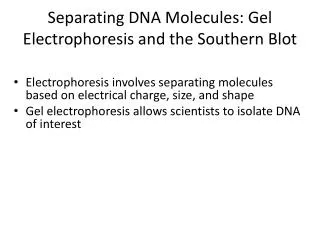
Analysis of DNA • Gel electrophoresis through agarose or poly-acrylamide gels is the most common method used to separate, identify and purify DNA fragments. DNA fragments are negatively charged molecules at neutral pH. Therefore, they will migrate towards the (+) electrode in an electric field. Both types of gels (agarose or poly-acrylamide) are: -porous -function as a molecular sieve -thereby separate DNAs based on SIZE.
Poly-acrylamide gels • Poly-acrylamide gels are most effective for separating small fragments of DNA (less than 1000 base pairs). • Their resolving power is extremely high, and fragments of DNA that differ in size by as little as 1 base pair can be separated from one another.
Agarose gels • More porous agarose gels have a lower resolving capacity than polyacrylamide gels. • Agarose gels however have a greater range of separation. • DNAs from 100 base pairs to approximately 50 kilo base pairs in length can be separated on agarose gels. • Most commonly used in the laboratory.
Properties of DNA and RNA molecules for gel fractioning • Both gels can be used to fractionate single-stranded DNA (or RNA molecules) by incorporating chemical denaturants in the gels.
Properties of denaturants: • Frequently used denaturants include: urea formamide formaldehyde Denaturants compete with and disrupt the hydrogen bonding between base pairs
Visualization of DNA and RNA • Fragments of DNA/RNA can be directly visualized using ethidium bromide (EtBr). • This toxic compound intercalates between the stacked bases of DNA/RNA and fluoresces when UV light is directed onto the gel. • Note: all nucleic acids (DNA and/or RNA) present in a gel are visualized and there is no discrimination based upon sequence.
Visualization of DNA with EtBr (C) (A) (B) • Gels are prepared with EtBr prior the migration of DNA/RNA samples. • After Gel electrophoresis (A) fragments of DNA/RNA can be directly visualized under UV light (B) and (C)
Southern Blotting • The technique was developed by E.M. Southern in 1975. The Southern blot is used to detect the presence of a particular DNA fragment in a sample. The DNA detected can be a single gene, or it can be part of a larger piece of DNA such as a viral genome.
Southern Blotting Probes: • Most commonly, the probe is radioactive. Other approaches include chemiluminescent compounds and fluorophores. • In the case of a radioactive probe, the membrane is exposed to X-ray film (autoradiography) and the film darkens where there is a concentration of radioactivity. • Chemiluminescent and fluorophores require specific wavelengths of light for detection.
Southern Blotting • Southern blot hybridization can be used to follow the inheritance of selected genes. • Because the genomic DNA samples are fractionated by separation of restriction fragments according to size, mutations which alter a restriction site, and significantly large deletions or insertions occurring between neighboring restriction sites, can be detected. • Such mutations will change the size of restriction fragments and the positions of bands in Southern blot analysis. This is called Restriction Fragment Length Polymorphism (RFLP).
RFLPRestriction Fragment Length Polymorphism • The existence of two or more variants at significant frequencies in the population is called polymorphism. • RFLP has been widely used in direct detection of disease-causing mutation, DNA fingerprinting, and linkage of polymorphism with gene mutation.
RFLPs commonly result from two types of DNA variants: Base changes in nucleotide sequence abolish existing restriction sites or create new restriction sites. Upon cleavage with restriction enzyme, a different band pattern (change in size) will be revealed in Southern blot hybridization. The change in size detected on the gel will be dependent on the effect of the base change, i.e. if a new site is created or a site is abolished.
Example of RFLP: Sickel cell Mutation Sickel cell mutation: The restriction enzyme Mst II recognizes and cleaves at the sequence CCTNAGG (where N is any nucleotide). The A to T mutation within the codon 6 of the -globin gene eliminates a cleavage site for the enzyme and generates a disease-specific RFLP.
Fluorescence in situ Hybridization (FISH) • FISH - a process which stains chromosomes or portions of chromosomes with fluorescent molecules Figure : chromosomes in metaphase stained with the fluorescent molecule DAPI
USE of FISH: to identify the presence and location of a region of DNA or RNA within chromosome preparations, fixed cells or tissue sectionsMEDICAL USE of FISH:to Identify chromosomal abnormalities; analysis of chromosome structural aberrations and ploidy determination.gene mapping studies, toxicological studies.
Advantage of FISH: • less labor-intensive method for confirming the presence of a DNA segment within an entire genome than other conventional methods like Southern blotting
FISH PROCEDURE: 1. Denature the chromosomes2. Denature the probe 3. Hybridization 4. Fluorescence staining 5. Examine slides or store in the dark 2 4 3 5 1
(or, Jumping Genes) TransposableElements For the first half of the 20th century, genes were seen as objects with fixed positions on the chromosomes. However, in the 1950s, Barbara McClintock showed in maize that certain DNA fragments, termed transposons, can be activated to "jump" from one position on a chromosome to another. She identified a particular chromosome breakage event that always occurred at the same locus on maize chromosome 9, which she named the "Ds" or "dissociation" locus. Barbara McClintock was awarded Nobel Prize in Physiology or Medicine, in the 1983
TransposableElements The plants containing a broken chromosome 9, showed stripes and spots of color, an indication that mutations had taken place in the developing body of the plant.
Philadelphia (Ph) chromosome Philadelphia (Ph) chromosome triggers a cancerous disease called Chronic Myeloid Leukemia (CML)
Leukemia is characterized by hyper-proliferation of immature white blood cells normalperson Leukemic patient red blood cells whiteblood cell hyperproliferation of white blood cells
Discovery of Philadelphia (Ph) chromosome In 1960, Peter Nowell’swork at the University of Pennsylvania School of Medicine, together with the late David Hungerford from the Fox Chase Cancer Center’s Institute for Cancer Research, established that patients with a form of leukemia had cells containing an abnormally small chromosome, soon named the Philadelphia chromosome
The Philadelphia (Ph) chromosome derives from translocation of a segment of the Chr. 9 to the Chr. 22. In normal individual Chr. 9 carries ablgene while Chr. 22has the bcr gene. 9; 22 Translocation fuses bcrand ablgenes. Leukemic patient normal individual bcr Bcr-abl Chr. 22 abl Chr. 9 De Klein et al. Nature 300, 765 (1982) Groffen et al. Cell 36, 93 (1984)
G 2 S M G 1 G0 BCR-Abl fusion affects multiple cell functions Proliferation & differentiation Stem cell turnover BCR-Abl Adapted from Jörgensen, 2001. Hem. Onc. Cytoskeleton/ adhesion defects Apoptosis
Fluorescence In Situ Hybridization (FISH) a tool for diagnosing CML fusion 22 bcr/abl fusion 9 abl/bcr abl bcr
Retinoblastoma is a cancerous disease in child Typically presents in first 2-3 years of life. Retinoblasts fail to differentiate - continue to divide, forming tumors in the retina. Retinoblastoma diplays a condition called Leukocoriaor “white pupil”
Retinoblastoma is caused by recessive mutation of retinoblastoma protein (RB1) localized on the Chr 13
RB1 function: in normal cell the retinoblastoma protein regulates the cell cycle Cell cycle = OFF Rb binds to E2F: no transcription, no entry into S phase Cell cycle = ON Rb does not bind to E2F: transcription and entry into S phase Mutation of RB1: no cell cycle arrest, cells proliferate indefinitely, can induce Cancer
FISH analysis of RB1 gene. Horizontal red and green bars indicate the regions covered by the probes The RB1 probe (red) spans the entire gene while the D13S1009 probe (green) hybridizes to the region surrounding the D13S1009 locus marker and serves as a control Figure 1). Figure 1 In normal diploid interphase nuclei and metaphase chromosomes, the probe generates two red and two green signals corresponding to the two normal chromosomes 13 (Figure 2). In cells with interstitial deletion of chromosome 13, in which the RB1 locus is deleted and the D13S1009 locus is retained, one red (RB1) and two green (D13S1009) signals will be observed (Figure 3). Figure 3 Figure 2
FISH and Telomeres • Special telomeric probes specific to individual chromosomes have been designed • Probe is based on the TTAGGG repeat present on all human telomeres
FISH and Telomeres • Application in cytogenetics - can detect submicroscopic deletions and cryptic translocations of genes associated withunexplained mental retardation and miscarriages
Analysis of RNA Gel electrophoresis through agarose gel RNA fragments are negatively charged molecules at neutral pH. Therefore, they will migrate towards the (+) electrode in an electric field.
Northern Blotting The technique was developed by Alwine and his colleagues in 1979 Technique for detecting specific RNAs separated by electrophoresis by hybridization to a labeled DNA probe.
Northern Blotting RNA Gel electrophoresis Transfer to membrane Probed with complimentary cDNA Autoradiography
Analysis of RNA • RNA especially has secondary structure that impedes its migration. • Agarose gels can be used to fractionate single-stranded RNA molecules by incorporating chemical denaturants in the gels. • Required step prior to resolution of RNA on the gel of choice.
Uses for Northern Blots? • 1. Temporal expression of genes. • 2. Spatial expression of genes • 3. Tissue specific expression of genes • 4. Splicing isoforms of genes
Detection of Proteins Coomassie Stain: 0.5 ug protein. Silver Stain: 0.25 ng protein. Resolved through polyacrylamide gels with SDS.
Western Blotting Protein Gel electrophoresis Transfer to membrane Probed with antibody Chromogenic reaction
Western Blotting 1 step : Gels are usually made by pouring them between two glass or plastic plates, using the gel solution. Western blot uses two different types of agarose gel: stacking and separating gel. The higher, stacking gel is slightly acidic (pH 6.8) and has a lower acrylamide concentration making a porous gel, which separates protein poorly but allows them to form thin, sharply defined bands. The lower gel, called the separating, or resolving gel, is basic (pH 8.8), and has a higher polyacrylamide content, making the gel's pores narrower.
Western Blotting 2 step : The samples are loaded into the wells, and the empty wells are loaded with sample buffer. The gel is then connected to the power supply and allowed to run. The proteins when loaded on the gel have a negative charge, as they have been denatured by heating, and will travel toward the positive electrode when a voltage is applied