Download
1 / 25
250 likes | 461 Views
The Endocrine System. Chapter 18. Introduction. Endocrine organs are ductless, well-vascularized glands that release hormones directly into the bloodstream or lymph. They are small and widely separated in the body.

E N D
The Endocrine System Chapter 18
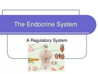
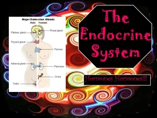
Introduction • Endocrine organs are ductless, well-vascularized glands that release hormones directly into the bloodstream or lymph. • They are small and widely separated in the body. • The major endocrine organs are the pituitary, thyroid, parathyroid, adrenal, pineal and thymus as well as pancreas an gonads. Hypothalamus is a neuroendocrine organ. • Hormonally regulated processes include reproduction, growth and development, mobilization of body defenses, maintenance of electrolyte, water and nutrient balance, and regulation of cellular metabolism. • Most hormones are steroids or amino acid derivatives.
Mechanisms of Hormone Action • Hormones alter cell activity by stimulating or inhibiting characteristic cellular processes. • Cell responses to hormone stimulation may involve changes in membrane permeability, enzyme synthesis, activation or inhibition, secretory activity, and gene activation. • Steroid hormones and thyroid hormone enter their target cells and effect responses by activating DNA, initiating mRNA formation that leads to protein synthesis.
Mechanism of Action • Second-messenger mechanisms by employing intracellular messengers and transduced by G proteins . • In the cAMP system, the hormone binds to a plasma membrane receptor coupled to adenylate cyclase which catalyzes the synthesis of cAMP from ATP. • cAMP initiates reactions that activate protein kinases and other enzymes. • Other messengers are PIP, cGMP and Calcium.
Hormone-Target Cell Specificity • The ability of a target cell to respond to a hormone depends on the presence of receptors, within the cell or on the plasma membrane to which the hormone can bind. • Hormone receptors are dynamic structures. Changes in number or sensitivity of receptors may occur in response to high or low levels of stimulating hormones.
Half-life, Duration and Control of Hormone Release • Hormone half-life and duration of activity are limited and vary from hormone to hormone. • Blood levels of hormone reflect a balance between secretion and degradation/excretion . The liver and kidneys are major organs that degrade hormones. Excretion is in feces or urine. • The endocrine organs are activated to release their hormones by humoral, neural or hormonal stimuli. • Negative feedback-controls levels in the blood. • The nervous system, acting through hypothalamic controls can override or modulate hormonal effects.
Major Endocrine Organs • The pituitary gland (Hypophysis) • this hangs from the base of the brain and is enclosed by bone. It consists of a hormone-producing glandular portion (ant.pit.) and a neural portion(post.pit.), which is an extension of hypothalamus. • Hypothalamus functions: regulates the hormonal output of the ant.pit.via rel.and inh.hormones and synthesizes two hormones that it exports to post.pit. For storage and later release. • Four of the six adenohypophseal hormones are tropic hormones that regulate the function of other endocrine organs.
Adenohypophseal Hormones • The ant.pit.divided into the pars distalis, pars intermedia and pars tuberalis. • The hypophyseal portal system ensures that all the blood entering the portal vessels will reach the intended target cells before it returns to gen. Circulation. • GH is an anabolic hormone that stimulates growth of all body tissues but especially skeletal muscle and bone. It mobilizes fats, stimulates protein synthesis and inhibits glucose uptake and metabolism. Secretion is regulated by GHRH (somatocrinin) and GHIH (somatostatin). Hypersecretion causes gigantism in children and acromegaly in adults. Hyposecretion in children causes pituitary dwarfism.
Adenohypophyseal Hormones • TSH promotes normal development and activity of the thyroid gland. TRH stimulates its release. Negative feedback of thyroid hormone inhibits it. • ACTH stimulates the adrenal cortex to release corticosteroids. ACTH release is triggered by CRH and inhibited by feedback inhibition of rising glucocorticoid levels.
Adenohypophyseal Hormones • The gonadotropins, FSH and LH regulate the function of gonads in both sexes. FSH: stimulates sex cell production. LH: stimulates gonadal hormone production. Gonadotropin levels rise in response to GnRH. Negative feedback of gonadal hormones inhibits gonadotropin release. • Prolactin (PRL) promotes milk production in humans. Controlled by PRH and PIH. • In many nonhuman vert. MSH stimulates melanocytes to produce melanin.
Neurohypophyseal Hormones • This contains the axons of the hypothalamic neurons. Neurons of the supraoptic and paraventricular nuclei manufacture ADH and oxytocin.. • Oxytocin stimulates powerful uterine contractions which trigger labor and delivery of infant and milk ejection. Also promotes sexual arousal and nurturing. Positive feedback. • ADH stimulates kidney tubules to reabsorb and conserve water. It is released in response to high solute concentration in the blood and inhibited by low solute concentration. Hyposecretion results in diabetes insipidus.
The Thyroid Gland • The thyroid gland is located in the anterior throat. The thyroid follicles store thyrglobulin, a colloid form of the hormone. • TH includes Thyroxine (T4) and triiodothyronine (T3), which increases the rate of cellular metabolism. • Secretion of thyroid hormone is prompted by TSH. • The follicle cells reuptake the stored colloid and split the hormone from the colloid for release.
The Thyroid Gland • Most T4 is converted to T3 in the target tissues. These hormones act on a steroid-like mechanism. • These hormones exert a calorigenic effect, which enables us to adapt to cold temps. • Hypersecretin-Graves’disease. Hyposecretion:cretinism in infants and myedema in adults. • Calcitonin-parafollicular (C) cells of the gland. Released in response to rising calcium levels. Depresses blood calcium levels by inhibiting bone matrix and enhancing calcium deposit in bone.
The Parathyroid Glands • These are located on the dorsal aspect of the thyroid gland. The chief cells secrete PTH. • PTH is the antagonistic of calcitonin. It causes an increase in calcium levels in the blood by targeting the bone, intestines and kidneys. • Triggered by falling blood calcium levels. • Hypersecretion-hypercalcemia. • Hyposecretion-hypocalcemia, tetany and respiratory paralysis.
The Adrenal Glands • The paired adrenal (suprarenal) glands sit atop the kidneys. Each adrenal gland has two functional parts-outer cortex and inner medulla. • Three groups of steroid hormones are produced by the cortex from cholesterol- mineralocorticoids, glucocorticoids and gonadocorticoids. • The adrenal medulla produces catecholamines-E and NE- in response to SNS stimulation. Its catecholamines enhance and prolong the fight-or-flight response to short-term stressors.
Adrenal Cortex Hormones • Mineralocorticoids (zona glomerulosa)-primarily aldosterone regulate sodium ion resorption by the kidneys and thus indirectly regulate levels of other electrolytes that are coupled to sodium transport. Release is stimulated by the renin-angitensin mechanism, rising potassium or sodium levels in the blood, and ACTH. • Glucocorticoids (zona fasciculata)-primarily cortisol are important metabolic hormones that help the body resist stressors by inc.blood glucose, fatty acid and amino acid levels and BP. High levels of this-depress the immune system and inflammatory response. ACTH -major stimulus for release.
Adrenal Cortex Hormones • Gonadocorticoids (zona reticularis)-mainly androgens are produced in small amounts throughout the life. • Hypoactivity of the adrenal cortex-Addison’s disease. • Hyperactivity-aldosteronism, Cushing’s disease, and/or masculinization. • Hyperactivity of medulla-symptoms typical of SNS overactivity.
The Pancreas • The pancreas, located in the abdomen close to the stomach is both an exocrine and endocrine gland. The endocrine portion (pancreatic islets) releases insulin and glucagon (plus pancreatic polypeptide and somatostatin-F and delta cells resp.) to the blood. • Glucagon-by cells when blood glucose levels are low, stimulates the liver to release glucose to the blood. • Insulin-by cells when blood glucose (and amino acids)levels are high. Increases uptake and metabolism. • Hyposecretion of insulin-diabetes mellitus. Signs are polyuria, polydipsia and polyphagia.
The Gonads • The ovaries of the female, located in the pelvic cavity, release two main hormones. • Secretion of estrogens by the ovarian follicles begins at puberty under the influence of FSH. They stimulate maturation of the female reproductive system and development of secondary sex characteristics. Progesterone is releasedin response to high blood levels of LH. It works with estrogens in establishing the menstrual cycle.
The Male Gonads • These are the testes located in the outside. • The testes of the male begin to produce testosterone at puberty in response to LH. • Testosterone promotes maturation of the male reproductive organs, development of secondary sex characteristics, and production of sperm by the testes.
The Pineal Gland and Thymus • This is located in the diencephlaon and its primary hormone is melatonin (pinealocytes), which influences daily rhythms and may antigonadotropic effects in humans. • The thymus gland, located importqant in the upper thorax, declines in size and function with age. Its hormones thymosins and thymopoietins are important to the normal development of the immune response.
The Kidneys • Endocrine cells in the kidneys produce calcitrol, erythropoietin and enzyme renin. • Calcitrol-stimulates Ca and PO ion absorption along GI tract. • EPO-stimulates red blood cell production by the bone marrow. • Renin-converts angiotensinogen to angiotensin I. To angiotensin II in lungs. This in turn stimulates production of aldosterone and ADH. Promotes thirst and vasoconstriction.
Other Hormone Producing Structures • Many body organs not normally considered endocrine contain isolated cell clusters that secrete hormones. • Examples include the heart-atrial natriuretic peptide; GI tract organs (gastrin, secretin, and others);the placenta (hormones of pregnancy-estrogen, progesterone and others);the kidneys (erythropoietin);and skin (cholecalciferol).
Patterns of Hormonal Interaction • Antagonistic-opposing effects • Synergistic-additive effects • Permissive-one hormone is necessary for another to produce its effect. • Integrative-different but complementary effects.
Other Factors • Growth-GH, thyroid, insulin, PTH, calcitrol, and gonadal hormones. • Stress-GAS. The alarm phase, the resistance phase and exhaustion phase. • Behavior-affect nervous system producing changes in mood, emotional stress and behavior. • Aging-decline in concentration of reproductive hormones.