Download
1 / 1
10 likes | 140 Views
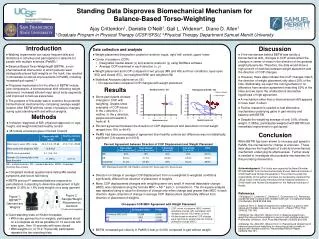
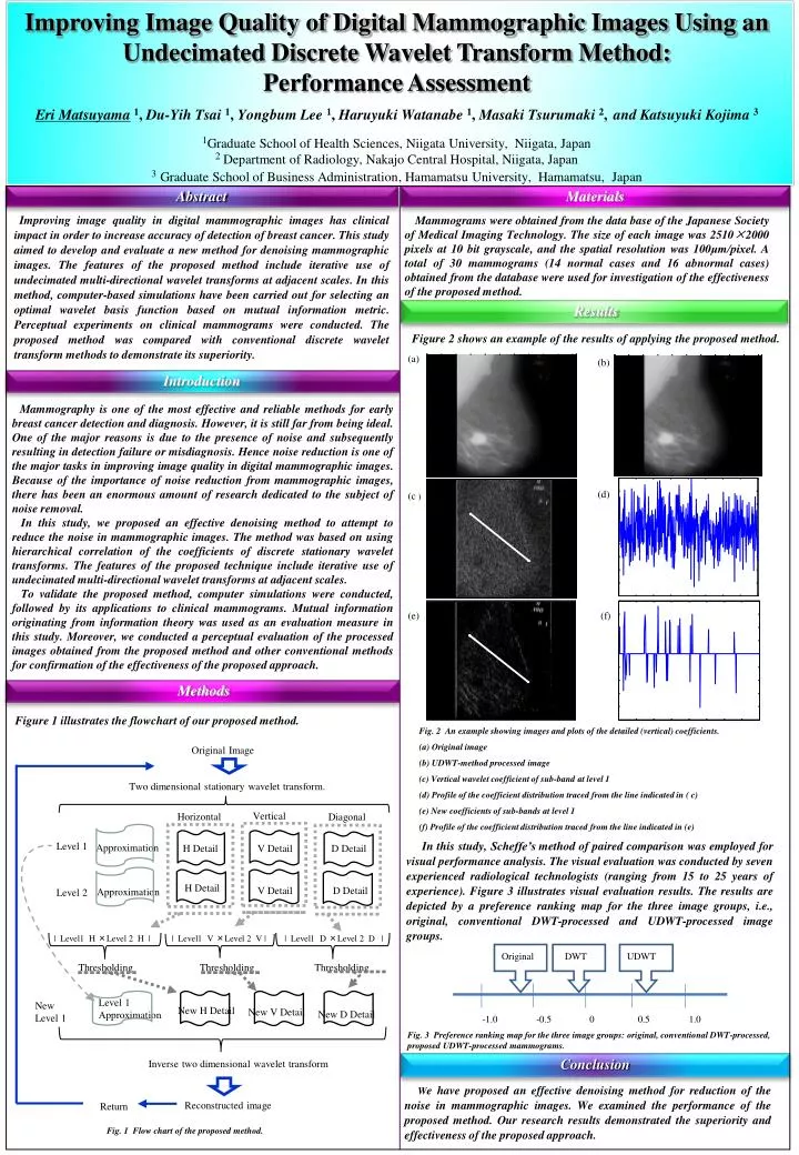
Improving Image Quality of Digital Mammographic Images Using an Undecimated Discrete Wavelet Transform Method: Performance Assessment Eri Matsuyama 1 , Du-Yih Tsai 1 , Yongbum Lee 1 , Haruyuki Watanabe 1 , Masaki Tsurumaki 2 , and Katsuyuki Kojima 3

E N D
Improving Image Quality of Digital Mammographic Images Using an Undecimated Discrete Wavelet Transform Method: Performance Assessment Eri Matsuyama1, Du-Yih Tsai 1, Yongbum Lee 1, Haruyuki Watanabe 1, Masaki Tsurumaki 2,and Katsuyuki Kojima 3 1Graduate School of Health Sciences, Niigata University, Niigata, Japan 2Department of Radiology, Nakajo Central Hospital, Niigata, Japan 3Graduate School of Business Administration, Hamamatsu University, Hamamatsu, Japan Original Image Abstract Materials Improving image quality in digital mammographic images has clinical impact in order to increase accuracy of detection of breast cancer. This study aimed to develop and evaluate a new method for denoising mammographic images. The features of the proposed method include iterative use of undecimated multi-directional wavelet transforms at adjacent scales. In this method, computer-based simulations have been carried out for selecting an optimal wavelet basis function based on mutual information metric. Perceptual experiments on clinical mammograms were conducted. The proposed method was compared with conventional discrete wavelet transform methods to demonstrate its superiority. Mammograms were obtained from the data base of the Japanese Society of Medical Imaging Technology. The size of each image was 2510×2000 pixels at 10 bit grayscale, and the spatial resolution was 100μm/pixel. A total of 30 mammograms (14 normal cases and 16 abnormal cases) obtained from the database were used for investigation of the effectiveness of the proposed method. Two dimensional stationary wavelet transform. Vertical Horizontal Diagonal Level 1 Approximation D Detail H Detail V Detail Results Figure 2 shows an example of the results of applying the proposed method. (a) H Detail (b) V Detail D Detail Approximation Level 2 Introduction Mammography is one of the most effective and reliable methods for early breast cancer detection and diagnosis. However, it is still far from being ideal. One of the major reasons is due to the presence of noise and subsequently resulting in detection failure or misdiagnosis. Hence noise reduction is one of the major tasks in improving image quality in digital mammographic images. Because of the importance of noise reduction from mammographic images, there has been an enormous amount of research dedicated to the subject of noise removal. In this study, we proposed an effective denoising method to attempt to reduce the noise in mammographic images. The method was based on using hierarchical correlation of the coefficients of discrete stationary wavelet transforms. The features of the proposed technique include iterative use of undecimated multi-directional wavelet transforms at adjacent scales. To validate the proposed method, computer simulations were conducted, followed by its applications to clinical mammograms. Mutual information originating from information theory was used as an evaluation measure in this study. Moreover, we conducted a perceptual evaluation of the processed images obtained from the proposed method and other conventional methods for confirmation of the effectiveness of the proposed approach. | Level1 H ×Level 2 H | | Level1 V ×Level 2 V | | Level1 D ×Level 2 D | Thresholding Thresholding Thresholding (d) (c ) Level 1 Approximation New Level 1 New H Detail New V Detail New D Detail (a) Inverse two dimensional wavelet transform (e) (f) Reconstructed image Return Methods Figure 1 illustrates the flowchart of our proposed method. Fig. 2 An example showing images and plots of the detailed (vertical) coefficients. (a) Original image (b) UDWT-method processed image (c) Vertical wavelet coefficient of sub-band at level 1 (d) Profile of the coefficient distribution traced from the line indicated in ( c) (e) New coefficients of sub-bands at level 1 (f) Profile of the coefficient distribution traced from the line indicated in (e) In this study, Scheffe’s method of paired comparison was employed for visual performance analysis. The visual evaluation was conducted by seven experienced radiological technologists (ranging from 15 to 25 years of experience). Figure 3 illustrates visual evaluation results. The results are depicted by a preference ranking map for the three image groups, i.e., original, conventional DWT-processed and UDWT-processed image groups. Fig. 3 Preference ranking map for the three image groups: original, conventional DWT-processed, proposed UDWT-processed mammograms. Conclusion We have proposed an effective denoising method for reduction of the noise in mammographic images. We examined the performance of the proposed method. Our research results demonstrated the superiority and effectiveness of the proposed approach. Fig. 1 Flow chart of the proposed method.