Download
1 / 51
570 likes | 932 Views
Response Evaluation of Gastrointestinal Stromal Tumors (GIST). Haesun Choi, M.D. Diagnostic Imaging The University of Texas MD Anderson Cancer Center, Houston, TX. Gastrointestinal Stromal Tumor (GIST). “KIT” receptor. Kinase domains. Tyrosine kinase receptor blocker.

E N D
Response Evaluation of Gastrointestinal Stromal Tumors (GIST) Haesun Choi, M.D. Diagnostic Imaging The University of Texas MD Anderson Cancer Center, Houston, TX
“KIT” receptor Kinase domains Tyrosine kinase receptor blocker Imatinib mesylate Chris Corless, M.D. +
“Computed Tomography (CT) and Magnetic Resonance Imaging (MRI) are the best currently available and most reproducible methods for measuring the target lesions …” Thessasse et al. JNCI 92(3); 205, 2000
Fluorine-18-fluorodeoxyglucose Positron Emission Tomography (FDG PET)
8/9/02 10/28/02
Pre-Treatment Pre-Treatment
Gastric GIST Metastatic GIST
Small bowel GIST Metastatic GIST
6/01 HU 63 3.3 cm 8/01 HU 38 2.3 cm 10/01 HU 32 1.9 cm
Pre-Treatment 2 Months Post
Pre-Treatment 5 Days Post
30 HU 43 HU Pre-Treatment 2 Months Post
Total patients = 36 CT* and PET* = 29 *within a week of each other Total lesions = 173 Liver: 116 Peritoneum: 52 Pleura: 5 Methods and Materials (I) • CT vs. PET PET: EORTC1999 • Tumor size (cm) • Tumor density (HU) • “Overall tumor status (OTS)”
Pre-Treatment Subjective Tumor Response Evaluation: OTS Size + • tumor vessels • solid tumor nodules • tumor density
Pre-Treatment 2 Months Post
Mean HU Size P<0.0025, t-test P = 0.0025, t-test Mean SUVmax P = 0.0025, t-test Objective Tumor Response Evaluation Pre-treatment 8 Wks Post-treatment
Size vs. SUV Note. - The data were analyzed for the 29 patients who underwent both CT and FDG PET. * Based on RECIST ** Based on modified EORTC 1999 criteria
Total patients = 40 CT and PET “Good Response” :Decrease in SUVmax >70% <2.5 Good Response:33 (83%) 30 (75%): PET CR 3 (8%): 70 - 99% decrease, decrease to a value <2.5 Poor Response:7 (17%) 5 (12%): stable 2 (5%): increased SUVmax Methods and Data Analysis (II) (Van den Abbeele AD, et al, ASCO 2002)
Changes in Size and HU on CT vs. Tumor Response on FDG PET Total number of patients = 40 n – number of patients
1 1 .9 .9 .8 .8 .7 .7 .6 .6 .5 .5 .4 .4 .3 .3 .2 .2 .1 .1 0 0 0 0 3 3 6 6 9 9 12 12 15 15 18 18 21 21 24 24 27 27 30 30 Modified CT Criteria PET response: SUV < 2.5, 70% CT response: HU -15%, Size -10% + + + + + + + + + + + + + + + + + + + + + + + + + + + + + P = 0.03 + + + + + + + + + + + + + + + + + + + + + + + + + + + + + + + + + + + + + + + + + + + P = 0.03 + + + + + Responder Responder Non-responder Non-responder Months Months Time to Progression by PET and modified CT criteria
Time to Progression: RECIST N=98 P = 0.1 Response Rate 45%
Time to Progression: Modified CT N=98 P = 0.0002 Response Rate 83%
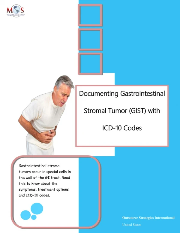
Progression • Increase in tumor size • Appearance of a new lesion at the site of primary tumor • Appearance metastatic lesions
Pre-Treatment 2 Months Post 8 Months Post 11 Months Post
10 Months Post 17 Months Post 21 Months Post 27 Months Post
Progression in GIST • “Increase in tumor size” • Appearance of a new lesion at the site of primary tumor • Appearance metastatic lesions “Appearance of new intra-tumoral nodules”
30 HU 43 HU Pre-Treatment 2 Months Post
Conclusions • RECIST underestimates the tumor response. • Subjective evaluation using changes in tumor nodules, density, tumor vessels, in addition to change in size is the best criteria on CT and is reproducible. • CT density alone can be a good indicator in early, quantitative tumor response evaluation.
Conclusions • Objective evaluation usinga combination of tumordensity (15% change) and modified tumor size criteria (10% change) is promising in early tumor response evaluation and has a prognostic value. • FDG PET should be performed whenever the CT findings are inconclusive or inconsistent with the clinical presentation.
Division of Diagnostic Imaging: Chusilp Charnsangavej, M.D. Silvana C. Faria, M.D. Eric P. Tamm, M.D. Evelyn M. Loyer, M.D. Kazama Toshiki, M.D. Division of Nuclear Medicine: Donald A. Podoloff, M.D. Homer A. Macapinlac, M.D. Department of Sarcoma Medical Oncology: Robert S. Benjamin, M.D. Sarcoma Center Team Department of Biostatistics: Marcella M. Johnson, M.S. Acknowledgements
Data Analysis: CT **OTR – overall tumor response *JNCI 92(3); 205, 2000
OTS vs. SUV P = 0.0001*, Chi-Square Test *Statistically significant. Note. - The data were analyzed for the 29 patients who underwent both CT and FDG PET.
HU vs. SUV P = 0.3088, Chi-Square Test Note. - The data were analyzed for the 29 patients who underwent both CT and FDG PET.
Methods and Materials (II) • Two radiologists who were not participated in initial analysis of CT images • Overall Tumor Status (OTS) • The results of two radiologists were compared with each other.
Mean HU Size P < 0.0001, t-test P < 0.0001, t-test Mean SUVmax P < 0.0001, t-test Pre-treatment 8 Wks Post-treatment
Reader A vs. B P* = 0.0002, Chi-Square Test, rtau** =0.5782 *Statistically significant. ** Kendall’s Tau correlation. Note – Grades are based on OTR at 8 wks post-treatment.
OTS vs. SUV P = 0.0001*, Chi-Square Test *Statistically significant. Note. - The data were analyzed for the 35 patients who underwent both CT and FDG PET.
Pre-Treatment 2 Months Post EatoEaton 411286 24 Months Post 27 Months Post
Discrepancy(?): HU vs. SUVmax • Development of intratumoral hemorrhage • Definition of ROI • EORTC guideline
“KIT” Receptor + Tyrosine Kinase Receptor Blocker
Conclusions • RECIST underestimates the tumor response in GIST. • Subjective evaluation using changes in tumor nodules, density, tumor vessels, in addition to change in size is the best criteria on CT and is reproducible.