Download
1 / 17
170 likes | 288 Views
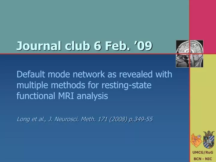
Journal club 6 Feb. ’09. Default mode network as revealed with multiple methods for resting-state functional MRI analysis Long et al., J. Neurosci. Meth. 171 (2008) p.349-55. Abstract.

E N D
Journal club 6 Feb. ’09 Default mode network as revealed with multiple methods for resting-state functional MRI analysis Long et al., J. Neurosci. Meth. 171 (2008) p.349-55
Abstract • Recently, human brain activity during a resting-state has attracted increasing attention. Several studies have found that there are two networks: the default mode network and its anti-correlation network. Some studies have subsequently showed that the functions of brain areas within the default mode network are crucial in human mental activity. To further discern the brain default mode network as well as its anti-correlation network during resting-state, we used three methods to analyze resting-state functional magnetic resonance imaging (fMRI) data; regional homogeneity analysis, linear correlation and independent component analysis, on four groups of dataset. Our results showed the existence of these two networks prominently and consistently during a resting- and conscious-state across the three methods. This consistency was exhibited in four independent groups of normal adults. Moreover, the current results provided evidences that the brain areas within the two anti-correlated networks are highly integrated at both the intra- and inter-regional level.
Materials & Methods • 4 sites with different scanners (Siemens/GE; 1.5-3.0T) • 4 x 10 healthy controls, matched across sites • 170 2-s EPI volumes, 3.4 mm in plane, 4-7 mm slices • Pre-processing: slice timing, realignment, normalization, resampling, temporal filtering (0.01-0.08 Hz), drift removal • Analyses • Regional homogeneity analysis (ReHo)local signal coherence • Functional connectivity analysis (SCA)long-range area-related coherence • Group independent component analysis (ICA)integrated network coherence • Conjunction analysiscombination
Take time courses si(tj) in a 3x3x3 voxel neighborhood For each time course, determine rank orders ri(tj) Calculate mean R(tj) over all time courses Calculate measure of deviation from expectation Kendall’s coefficient of concordance KCC = 0.015 KCC = 0.324 KCC = 0.432 KCC = 0.983 ReHo
ReHo • Individual smoothed KCC maps were divided by global mean • Group level one-tailed t-test to determine KCC > 1.0 (Pcorr < 0.05) A: posterior cingulate cortex B: medial prefrontal cortex C: angular gyrus D: supplementary motor area E: inferior parietal lobe F: insula G: medial temporal cortex H: inferior temporal cortex I: dorsolateral prefrontal cortex J: lingual/fusiform gyrus K: parahippocampal gyrus Group I Group II Group III Group IV
Seed ROI was defined in PCC (-5,-49,40) For each voxels, partial correlations were determined with 1 Seed ROI signal 6 Motion parameters 1 Global mean signal Seed ROI correlations were converted to z-values using Fisher transform Smoothing SCA
SCA • Group level two-tailed t-test to determine Z ≠ 0.0 (Pcorr < 0.05) • Significant positive correlations for pairs of anti-correlated areas A: posterior cingulate cortex B: medial prefrontal cortex C: angular gyrus D: medial temporal cortex E: inferior parietal lobe F: supplementary motor area G: insula H: dorsolateral prefrontal cortex I: inferior temporal cortex J: postcentral gyrus Group I Group II Group III Group IV
Principal component analysis extract 20 most important components Sphering decorrelates all components in spatial domain ICA rotation transforms decomposition into maximally independent components ICA
ICA • Default mode component was selected based on PCC activity • Individual independent components IC were backprojected • Group level two-tailed t-test to determine IC ≠ 0.0 (Pcorr < 0.05) A: posterior cingulate cortex B: medial prefrontal cortex C: angular gyrus D: inferior parietal lobe E: insula F: supplementary motor area G: medial temporal cortex H: inferior temporal cortex I: dorsolateral prefrontal cortex Group I Group II Group III Group IV
Conjunction analysis • Conjuncted (‘AND’) logical map was constructed from thresholded results A: posterior cingulate cortex B: medial prefrontal cortex C: angular gyrus D: inferior temporal cortex E: dorsolateral prefrontal cortex F: insula Group I Group II Group III Group IV
Discussion & Conclusion • Two anti-correlated networks with dynamic ongoing activity were found • Task-negative default mode network (PCC+MPFC+IPC) • Task-positive anti-correlated network (SMA+INS+DLPFC) • Multiple methods reveal strong synchronization • intra-regionally (ReHo) • inter-regionally (SCA+ICA) possibly for the purpose of efficient parallel processing
Discussion & Conclusion • Consistent findings across • Groups • Methods • Some differences were observed • ReHo more sensitive to task-negative than task-positive network • ReHo revealed areas outside both networks with unclear resting state function • SCA vs. ICA: area-related vs. network-related
Discussion & Conclusion • Limitations • Group differences • Thresholding • Physiological noise signals
Perspective • Default network • Marcus Raichle coined "default-mode" in 2001 to describe resting state brain function; the concept rapidly became a central theme in neuroscience. The default network is a network of brain regions that are active when the individual is not focused on the outside world and the brain is at wakeful rest. Also called the default mode network (DMN) or task-negative network (TNN), it is characterized by coherent neuronal oscillations at a rate lower than 0.1 Hz (one every ten seconds). During goal-oriented activity, the DMN is deactivated and another network, the task-positive network (TPN) is activated. It is thought that the default network corresponds to task-independent introspection, or self-referential thought, while the TPN corresponds to action, and that perhaps the TNN and TPN should be considered elements of a single default network with anticorrelated components. From: Wikipedia
Perspective • Anatomy • The default network is an interconnected and anatomically defined brain system that preferentially activates when individuals focus on internal tasks such as daydreaming, envisioning the future, retrieving memories, and gauging others' perspectives. It is negatively correlated with brain systems that focus on external visual signals. Its subsystems include part of the medial temporal lobe for memory, part of the medial prefrontal cortex for mental simulations, and the posterior cingulate cortex for integration, along with the adjacent precuneus and the medial, lateral and inferior parietal cortex. In the infant brain, there is limited evidence of the default network, but default network connectivity is more consistent in children aged 9-12 years, suggesting that the default network undergoes developmental change. From: Wikipedia
Perspective • Function • In humans, the default network has been hypothesized to generate spontaneous thoughts during mind-wandering and believed to be an essential component of creativity. It has been hypothesized to be relevant to mental disorders including Alzheimer's disease, autism, and schizophrenia. In particular, reduced default network activity has been associated with autism, overactivity with schizophrenia, and impaired control of entering and leaving the default network state is correlated with old age. The hypothesis that the default network is related to internally directed thought is not universally accepted. In 2007 the concept of the default mode was criticized as not being useful for understanding brain function, on the grounds that a simpler hypothesis is that a resting brain actually does more processing than a brain doing certain "demanding" tasks, and that there is no special significance to the intrinsic activity of the resting brain. From: Wikipedia
Perspective • Some overview papers • A default mode of brain function.Raichle et al. (2001). Proc Natl Acad Sci USA 98 (2): 676-82. • A default mode of brain function: a brief history of an evolving idea.Raichle et al. (2007). Neuroimage 37 (4): 1083-90. • Does the brain have a baseline? Why we should be resisting a rest.Morcom et al. (2007). Neuroimage 37 (4): 1073-82. • The brain's default network: anatomy, function, and relevance to disease.Buckner et al. (2008). Ann NY Acad Sci 1124: 1-38. • Spontaneous low-frequency blood oxygenation level-dependent fluctuations and functional connectivity analysis of the 'resting' brain.Auer (2008). Magn Reson Imaging 26 (7):1055-64. • Intrinsic brain activity in altered states of consciousness: how conscious is the default mode of brain function?Boly et al. (2008). Ann NY Acad Sci 1129:119-29. • The maturing architecture of the brain's default network.Fair et al. (2008). Proc Natl Acad Sci USA 105 (10):4028-32 • Default-mode brain dysfunction in mental disorders: a systematic review.Broyd et al. (2008). Neurosci Biobehav Rev (in press)