Download
1 / 48
580 likes | 1.19k Views
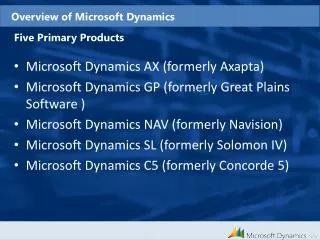
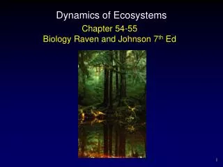
Overview of Dynamics. Judith Klein-Seetharaman Co-Course Director jks33@pitt.edu. Objectives of this Lecture. What is dynamics? Time scales of dynamics Methods to study dynamics What data do you get typically? Example: DNA binding. What is dynamics?. What is dynamics?. Definition:

E N D
Overview of Dynamics Judith Klein-SeetharamanCo-Course Director jks33@pitt.edu
Objectives of this Lecture • What is dynamics? • Time scales of dynamics • Methods to study dynamics • What data do you get typically? • Example: DNA binding Molecular Biophysics 3: Lecture 6
What is dynamics? Molecular Biophysics 3: Lecture 6
What is dynamics? • Definition: = changes in the position of the atoms in a molecule relative to each other or relative to an outside reference point. http://www.bioc.aecom.yu.edu/labs/girvlab/nmr/course/relaxdyn Proteins and other biological molecules are dynamic. Molecular Biophysics 3: Lecture 6
What are the time scales? Molecular Biophysics 3: Lecture 6
Example: Time-scales in rhodopsin Time-scales here from fs to min Molecular Biophysics 3: Lecture 6
Time scales of protein motions Overall tumbling Libration Slow loop reorientation Fast loop reorientation Vibration Side chain rotation/reorient. S-S flipping Aromatic ring flips 10-9 10-12 10-6 103 10 10-3 fs ps ns ms ms seconds minutes-hours-days ms-days: proton exchange ns-ps: fast internal motions ms-ms: slow internal motions Time scales from ps to days Molecular Biophysics 3: Lecture 6
What are the methods to study dynamics? Molecular Biophysics 3: Lecture 6
NMR parameters and time-scales Overall tumbling Librations Slow loop reorientation Fast loop reorientation Vibration Side chain rotation/reorient. S-S flipping Aromatic ring flips 10-9 10-12 10-6 103 10 10-3 fs ps ns ms ms seconds minutes-hours-days ms-days: proton exchange ns-ps: fast internal motions ms-ms: slow internal motions T2, T1r T1, T2, NOE HN exchange J Chemical shift Except to some degree in ms-ms range, NMR can report on all time-scales Molecular Biophysics 3: Lecture 6
Other methods and time-scales Overall tumbling Librations Slow loop reorientation Fast loop reorientation Vibration Side chain rotation/reorient. S-S flipping Aromatic ring flips 10-9 10-12 10-6 103 10 10-3 fs ps ns ms ms seconds minutes-hours-days ms-days: proton exchange ns-ps: fast internal motions ms-ms: slow internal motions Fluorescence IR, Raman HN exchange with mass spec Some biophysical measurements are fast… Molecular Biophysics 3: Lecture 6
Trapping of conformations Overall tumbling Librations Slow loop reorientation Fast loop reorientation Vibration Side chain rotation/reorient. S-S flipping Aromatic ring flips 10-9 10-12 10-6 103 10 10-3 fs ps ns ms ms seconds minutes-hours-days ms-days: proton exchange ns-ps: fast internal motions ms-ms: slow internal motions Light Rapid Mixing NMR, x-ray Some biophysical measurements take a long time… Molecular Biophysics 3: Lecture 6
Functions of dynamics and the time-scales Blue: types of motions Red: functional categorization Allosteric regulation / global conformational changes Ligand/protein binding Chemical kinetics Catalysis Overall tumbling Local folding global Libration Slow loop reorientation Fast loop reorientation Vibration Side chain rotation/reorient. S-S flipping Aromatic ring flips 10-9 10-12 10-6 103 10 10-3 fs ps ns ms ms seconds minutes-hours-days ms-days: proton exchange ns-ps: fast internal motions ms-ms: slow internal motions Internal motions are needed to provide flexiblity for functional motions. Molecular Biophysics 3: Lecture 6
Trp265 Ala169 Trp265 Ala169 Need for protein dynamics in rhodopsin? Dark – state structure Ligand: 11-cis retinal, no clashes Light-activated structure? Dark – state structure Ligand: all-trans retinal, steric clashes Molecular Biophysics 3: Lecture 6
Example: Function in Rhodopsin Definition and Function Example: Rhodopsin Function in Signal Transduction requires conformational changes Dark (inactive) Rhodopsin 11-cis retinal does not bind to G protein hn Light-activated Rhodopsin All-trans retinal does bind to G protein involves protein-ligand and protein-protein interactions Biomolecular motions are needed for function. Example for function: biomolecular interaction. Molecular Biophysics 3: Lecture 6
The type of data you can expect Molecular Biophysics 3: Lecture 6
Atomic resolution method - example X-ray Gives you snap shots of diffraction patterns in different states 2bcc 1bcc Zhang, Z., Huang, L., Shulmeister, V.M., Chi, Y.I., Kim, K.K., Hung, L.W., Crofts, A.R., Berry, E.A., Kim, S.H. Electron transfer by domain movement in cytochrome bc1. Naturev392pp.677-684 , 1998 Molecular Biophysics 3: Lecture 6
More snap shots 1up5 – calmodulin Ca-bound 1ctr – calmodulin free http://www.molmovdb.org/ http://www.bmb.psu.edu/faculty/tan/lab/gallery/calmodulin.jpg Molecular Biophysics 3: Lecture 6
Any problems? Molecular Biophysics 3: Lecture 6
Any problems?No information on time-scales. Molecular Biophysics 3: Lecture 6
Time-resolved spectroscopy Intrinsic Trp fluorescence Quenching by iodine Binding of ANS Dobson,et.al. 1994 Molecular Biophysics 3: Lecture 6
Any problems? Molecular Biophysics 3: Lecture 6
Any problems?Not atomic level information. Molecular Biophysics 3: Lecture 6
NMR parameters and time-scales Overall tumbling Librations Slow loop reorientation Fast loop reorientation Vibration Side chain rotation/reorient. S-S flipping Aromatic ring flips 10-9 10-12 10-6 103 10 10-3 fs ps ns ms ms seconds minutes-hours-days ms-days: proton exchange ns-ps: fast internal motions ms-ms: slow internal motions T2, T1r T1, T2, NOE HN exchange J Chemical shift Except to some degree in ms-ms range, NMR can report on all time-scales Molecular Biophysics 3: Lecture 6
Chemical Exchange http://www.oci.unizh.ch/group.pages/zerbe/NMR.pdf Molecular Biophysics 3: Lecture 6
H/D exchange can be measured in several ways • Slow exchange lifetimes (from mins to days) by following the loss of HN signal intensity of a protein dissolved in D2O. • Faster exchange lifetimes (5–500 ms) by following the exchange of HN magnetization with that of water protons. • At high pH directly measure the timescale of rate limiting conformational openings Molecular Biophysics 3: Lecture 6
HD Exchange and NMR Hernandez, G., Jenney, F.E.J., Adams, M.W. & LeMaster, D.M. Proc. Natl. Acad. Sci. USA 97, 3166–3170 (2000). Molecular Biophysics 3: Lecture 6
Relaxation and the NOE http://www.oci.unizh.ch/group.pages/zerbe/NMR.pdf • Longitudinal relaxation (T1): return of longitudinal (z-component) to its equilibrium value • Transverse relaxation (T2): decay of transverse (x,y-component) • Heteronuclear NOE Due to dipole interactions between different nuclei Molecular Biophysics 3: Lecture 6
From experiments to dynamics data Palmer, A.G., 3rd, M. Rance, and P.E. Wright,.J Amer Chem Soc, 1991 Molecular Biophysics 3: Lecture 6
Dynamics in folded/unfolded lysozyme Unfolded: Arrows indicate oxidized (all disulfide bonds present) lysozyme Folded: Molecular Biophysics 3: Lecture 6
NMR parameters and time-scales Overall tumbling Librations Slow loop reorientation Fast loop reorientation Vibration Side chain rotation/reorient. S-S flipping Aromatic ring flips 10-9 10-12 10-6 103 10 10-3 fs ps ns ms ms seconds minutes-hours-days ms-days: proton exchange ns-ps: fast internal motions ms-ms: slow internal motions T2, T1r T1, T2, NOE HN exchange J Chemical shift Except to some degree in ms-ms range, NMR can report on all time-scales Molecular Biophysics 3: Lecture 6
Amplitudes and Frequencies Molecular Biophysics 3: Lecture 6
Popular approach to quantify motions Measure R1, R2, heteronuclear NOE “model free” approach Get order parameter S2 ,τe, τm Molecular Biophysics 3: Lecture 6
Lipari-Szabo Model Free Approach http://www.oci.unizh.ch/group.pages/zerbe/NMR.pdf Molecular Biophysics 3: Lecture 6
Lipari Szabo http://www.oci.unizh.ch/group.pages/zerbe/NMR.pdf • Order parameters S2 • τe, effective correlation function time for internal motions • τm, overall tumbling correlation time for global motions Molecular Biophysics 3: Lecture 6
Lipari–Szabo “model-free” approach • Estimate (τm) from R2/R1 for a selected subset of the residues • fits to the observed relaxation data using various regression variables • model-selection criteria are used to decide which choice is appropriate for each residue • Reoptimize using the selected models. • Uncertainties in the optimized parameters were obtained by Monte Carlo simulation. Michael Andrec, Gaetano T. Montelione, RonaldM. Levy Journal of Magnetic Resonance 139, 408–421 (1999) Molecular Biophysics 3: Lecture 6
Example: DNA binding Molecular Biophysics 3: Lecture 6
Example: GCN4 Leucine Zipper Low S2 indicates high flexibility. S2 can be used to estimate energetics. Molecular Biophysics 3: Lecture 6
Energetic Components of Protein-DNA Interactions Adapted from Jen-Jacobson L., Biopolymers (1997) The observed free energy (green arrow) for specific binding is the net of large opposing energies. Molecular Biophysics 3: Lecture 6
Sources of Entropy and Enthalpy in Protein-DNA Interactions ∆Go=∆Ho–T∆So Molecular Biophysics 3: Lecture 6
INTER-DEPENDENT Strain energy Molecular Strain When atoms, functional groups or residues (sidechains, bases) adopt positions that are not their own positions of minimum potential energy Can result from: Bond bending Bond rotation Steric repulsion Electrostatic repulsion Strain energy: The energetic cost of strain Molecular Biophysics 3: Lecture 6
Thermodynamic parameters • H:Enthalpy measure of heat energy • S:Entropy measure of disorder • G: Gibbs Free Energy G H – TS • C: Heat capacity measure of the ability of a body to store heat • ∆CoP=(∂∆Ho/∂T)P =T(∂∆So/∂T)P Using ∆CoP we can calculate ∆Ho, ∆So, and ∆GTat any temperature. Molecular Biophysics 3: Lecture 6
Factors affecting ∆CoP free protein + specific DNA ↔ protein-DNA complex • ∆CoP is made more positive by: • • Burial and desolvation of polar surface • • Molecular strain • ∆CoP is made more negative by: • •Burial and desolvation ofnonpolar surface • •Losses of configurational/vibrational freedom • (Interface restrains sidechains, bases, backbone) • •Restricted freedom of interfacial H2O • •Linked equilibria (e.g. protonation, ion binding) Molecular Biophysics 3: Lecture 6
Proposal of Spolar & Record(1994) hydrophobic effect and conformational change Compared the measured heat capacity changes and the calculated changes in nonpolar ASA and polar ASA ∆CoP is much more negative than predicted. The “excess” ∆CoP could be accounted for by local folding coupled to binding. The observed ∆CoP and ∆So could be used to estimate the number of protein residues that fold upon DNA binding. Equation estimates -1.2 kJ K-1 mol-1 for DSconf “Conformational changes in the protein that buried large amounts of nonpolar surface are coupled to binding.” Spolar ad Record, Science (1994) Molecular Biophysics 3: Lecture 6
Example: GCN4 Leucine Zipper Low S2 indicates high flexibility. S2 can be used to estimate energetics. Molecular Biophysics 3: Lecture 6
From order parameter to entropy Sum of the order paramaters in bound form … in free form Result: DS = -0.6 kJ K-1 mol-1 • Calorimetric data estimates -1.2 kJ K-1 mol-1 • MD simulations suggest that 40-45% of total conformational entropy loss arises from backbone chain entropy, rest from side-chain • NMR result fits well with calorimetric experiment Molecular Biophysics 3: Lecture 6
Specific vs. non-specific complexesof the Lac-repressor Kalodimos et al, Science(2004) Molecular Biophysics 3: Lecture 6
Contacts of Lac repressor protein with nonspecific and specific DNA Kalodimos et al, Chem.Rev.(2004) “The same set of residues can switch roles from a purely electrostatic interaction with the DNA backbone in the nonspecific complex to a highly specific binding mode with the base pairs of the cognate operator sequence.” Molecular Biophysics 3: Lecture 6
Summary of this Lecture • Study of biomolecular interactions and dynamics are important to understand function of biomolecules. Biomolecular interactions Dynamics Function Molecular Biophysics 3: Lecture 6