Download
1 / 97
980 likes | 1.18k Views
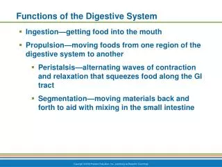
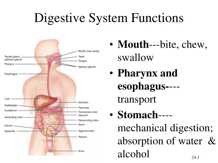
Digestive System Functions. Mouth ---bite, chew, swallow Pharynx and esophagus- ---transport Stomach ----mechanical digestion; absorption of water & alcohol. Small intestine- -chemical & mechanical digestion & absorption Large intestine- ---absorb electrolytes & vitamins (B and K)

E N D
Digestive System Functions • Mouth---bite, chew, swallow • Pharynx and esophagus----transport • Stomach----mechanical digestion; absorption of water & alcohol
Small intestine--chemical & mechanical digestion & absorption • Large intestine----absorb electrolytes & vitamins (B and K) • Rectum and anus---defecation
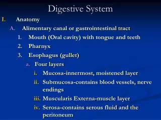
Layers of the GI Tract 1. Mucosal layer 2. Submucosal layer 3. Muscularis layer • Skeletal – in mouth, phayrnx, upper esophagus, and anus • control over swallowing and defecation • Smooth - mixes, crushes, and propels food along by peristalsis 4. Serosa layer- covers organs and walls of cavities not open to the outside
Peritoneum • Peritoneum • visceral layer covers organs • parietal layer lines the walls of body cavity • Peritoneal cavity • potential space containing a bit of serous fluid
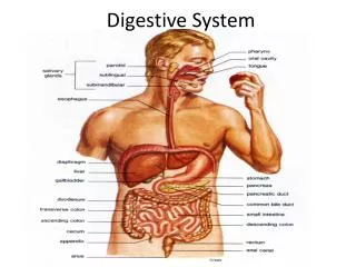
Salivary Glands • Parotid below your ear and over the masseter • Submandibular is under lower edge of mandible • Sublingual is deep to the tongue in floor of mouth
Composition and Functions of Saliva • Wet food for easier swallowing • Dissolves food for tasting • Buffers acidic foods • bulemia---vomiting hurts the enamel on your teeth • Chemical digestion of starch begins with enzyme (salivary amylase) • Enzyme (lysozyme) ---helps destroy bacteria • Protects mouth from infection with its rinsing action---1 to 1.5 qts/day
Composition of Teeth • Enamel • hardest substance in body • calcium phosphate or carbonate • Dentin • calcified connective tissue • Cementum • bone-like • periodontal ligament penetrates it What is the gingiva?
Dentition • Primary or baby teeth • 20 teeth that start erupting at 6 months • 1 new pair of teeth per month • Permanent teeth • 32 teeth that erupt between 6 and 12 years of age
Digestion in the Mouth • Mechanical digestion (mastication or chewing) • breaks into pieces • mixes with saliva so it forms a bolus • Chemical digestion • amylase • begins starch digestion at pH of 6.5 or 7.0
Esophagus • Collapsed muscular tube • In front of vertebrae • Posterior to trachea and heart • Pierces the diaphragm at hiatus
Physiology of the Esophagus - Swallowing • Voluntary phase---tongue pushes food to back of oral cavity • Involuntary phase----pharyngeal stage • breathing stops & airways are closed • soft palate & uvula are lifted to close off nasopharynx • vocal cords close • epiglottis is bent over airway as larynx is lifted
Anatomy of Stomach • Left side of body • Size when empty? • large sausage • stretches due to rugae- folds • Parts of stomach • Cardia – closest to esophagus • Fundus---top • Body • Pylorus---starts to narrow as approaches pyloric sphincter (toward small instestine) • Empties as small squirts of chyme leave the stomach through the pyloric valve
Muscularis • Three layers of smooth muscle-- outer longitudinal, circular & inner oblique • Permits greater churning & mixing of food with gastric juice
Mechanical Digestion • Gentle mixing waves • every 15 to 25 seconds • mixes bolus with 2 quarts/day of gastric juice to turn it into chyme (a thin liquid) • More vigorous waves • travel from body of stomach to pyloric region • Intense waves near the pylorus • open it and squirt out 1-2 teaspoons full with each wave
Chemical Digestion • Protein digestion begins • HCl denatures (unfolds) protein molecules • HCl kills microbes in food • Mucous cells protect stomach walls from being digested with 1-3mm thick layer of mucous
Absorption by the Stomach • Water especially if it is cold • Electrolytes • Some drugs (especially aspirin) & alcohol • Fat content in the stomach slows the passage of alcohol to the intestine where absorption is more rapid • Females have less total body fluid that same size male so end up with higher blood alcohol levels with same intake of alcohol
Cephalic Phase = “Stomach Getting Ready” • Cerebral cortex =sight, smell, taste & thought • stimulate parasympathetic nervous system
Gastric Phase = “Stomach Working” • Nervous control keeps stomach active • stretch receptors provide information • vigorous peristalsis and glandular secretions continue • chyme is released into the duodenum
Intestinal Phase = “Stomach Emptying” • Stretch receptors in duodenum slow stomach activity & increase intestinal activity • fatty acids or sugar signals medulla (in brain) • sympathetic nerves slow stomach activity
Anatomy of the Small Intestine • 20 feet long----1 inch in diameter • Large surface area for majority of absorption • 3 parts • duodenum---1 foot • jejunum---8 feet • ileum---12 feet • ends at ileocecal valve
Mechanical Digestion in the Small Intestine • Weak peristalsis in comparison to the stomach---chyme remains for 3 to 5 hours • Segmentation---local mixing of chyme with intestinal juices---sloshing back & forth
Absorption of Water • 9 liters of fluid dumped into GI tract each day • Small intestine reabsorbs 8 liters • Large intestine reabsorbs 90% of that last liter • Absorption is by osmosis through cell walls into capillaries inside villi
Anatomy of Large Intestine • 5 feet long by 2½ inches in diameter • Cecum & appendix • Rectum = last 8 inches of GI tract • Anal canal = last 1 inch of GI tract • internal sphincter----smooth muscle & involuntary • external sphincter----skeletal muscle & voluntary control
Mechanical Digestion in Large Intestine • Peristaltic waves (3 to 12 contractions/minute) • haustral churning----relaxed pouches are filled from below by muscular contractions (elevator) • gastroilial reflex = when stomach is full, gastrin hormone relaxes ileocecal sphincter so small intestine will empty and make room • gastrocolic reflex = when stomach fills, a strong peristaltic wave moves contents of transverse colon into rectum
Chemical Digestion in Large Intestine • No enzymes are secreted only mucous • Bacteria ferment • undigested carbohydrates into carbon dioxide & methane gas • undigested proteins into simpler substances-odor • turn bilirubin into simpler substances that produce color • Bacteria produce vitamin K and B in colon
Absorption & Feces Formation in the Large Intestine • Some electrolytes---Na+ and Cl- • After 3 to 10 hours, 90% of H2O has been removed from chyme • Feces are semisolid by time reaches transverse colon • Feces = dead epithelial cells, undigested food such as cellulose, bacteria (live & dead)
Defecation • Gastrocolic reflex moves feces into rectum • Stretch receptors signal sacral spinal cord • Parasympathetic nerves contract muscles of rectum & relax internal anal sphincter
Vomiting (emesis) • Forceful expulsion of contents of stomach & duodenum through the mouth • Cause: • Irritation or distension of stomach • Unpleasant sights, general anesthesia, dizziness, and certain drugs • Sensory input from medulla cause stomach contraction & complete sphincter relaxation • Serious because loss of gastric juices can lead to alkalosis
Diseases of the GI Tract • Dental caries and periodontal disease - tooth decay, gingivitis (in the tooth socket or peridontal ligament) – can lead to heart disease Cause: sugar and bacteria eat away enamel and dentin • Peptic Ulcers – lesion in stomach – bleeding is a problem, can become anemic Cause: • Bacteria – helicobactor pyloris • Aspirin – anit-inflammatory’s • Hypersecretions of HCl – from stress
Diverticulitis - sac-like pouch in intestine (like a hernia in the colon) – muscularis layer is weakened Symptoms: pain, increased defecation, nausea • Colorectoal Cancer - #2 leading cancer for men after lung cancer #3 for women after lung and breast cancer Symptoms: pollyps – pre-cancerous growths Prevention – GET SCREENED! Low fat diet and low intake of alcohol
Mucosa • Epithelium • stratified squamous(in mouth,esophagus & anus) = tough • simple columnar in the rest • secretes enzymes and absorbs nutrients • specialized cells (goblet) secrete mucous onto cell surfaces • enteroendocrine cells---secrete hormones controlling organ function • Lamina propria • thin layer of loose connective tissue • contains BV and lymphatic tissue • Muscularis mucosae---thin layer of smooth muscle • causes folds to form in mucosal layer • increases local movements increasing absorption with exposure to “new” nutrients
Submucosa • Loose connective tissue • containing BV, glands and lymphatic tissue • Meissner’s plexus--- • parasympathetic • innervation • vasoconstriction • local movement by muscularis mucosa smooth muscle
Muscularis • Skeletal muscle = voluntary control • in mouth, pharynx , upper esophagus and anus • control over swallowing and defecation • Smooth muscle = involuntary control • inner circular fibers & outer longitudinal fibers • mixes, crushes & propels food along by peristalsis • Auerbach’s plexus (myenteric)-- • both parasympathetic & sympathetic innervation of circular and longitudinal smooth muscle layers
Serosa • An example of a serous membrane • Covers all organs and walls of cavities not open to the outside of the body • Secretes slippery fluid • Consists of connective tissue covered with simple squamous epithelium
Parts of the Peritoneum • Mesentery • Mesocolon • Lesser omentum • Greater omentum • Peritonitis = inflammation • trauma • rupture of GI tract • appendicitis • perforated ulcer
Peritonitis • Acute inflammation of the peritoneum • Cause • contamination by infectious microbes during surgery or from rupture of abdominal organs
Pharyngeal Arches • Two skeletal muscles • Palatoglossal muscle • extends from palate to tongue • forms the first arch • posterior limit of the mouth • Palatopharyngeal muscle • extends from palate to pharyngeal wall • forms the second arch • behind the palatine tonsil
Salivary Gland Cellular Structure • Cells in acini (clusters) • Serous cells secrete a watery fluid • Mucous cells (pale staining) secrete a slimy, mucus secretion
Mumps • Myxovirus that attacks the parotid gland • Symptoms • inflammation and enlargement of the parotid • fever, malaise & sour throat (especially swallowing sour foods) • swelling on one or both sides • Sterility rarely possible in males with testicular involvement (only one side involved) • Vaccine available since 1967
Histology of the Esophagus • Mucosa = stratified squamous • Submucosa = large mucous glands • Muscularis = upper 1/3 is skeletal, middle is mixed, lower 1/3 is smooth • upper & lower esophageal sphincters are prominent circular muscle • Adventitia = connective tissue blending with surrounding connective tissue--no peritoneum
Swallowing • Upper sphincter relaxes when larynx is lifted • Peristalsis pushes food down • circular fibers behind bolus • longitudinal fibers in front of bolus shorten the distance of travel • Travel time is 4-8 seconds for solids and 1 sec for liquids • Lower sphincter relaxes as food approaches
Gastroesophageal Reflex Disease • If lower sphincter fails to open • distension of esophagus feels like chest pain or heart attack • If lower esophageal sphincter fails to close • stomach acids enter esophagus & cause heartburn (GERD) • for a weak sphincter---don't eat a large meal and lay down in front of TV • smoking and alcohol make the sphincter relax worsening the situation • Control the symptoms by avoiding • coffee, chocolate, tomatoes, fatty foods, onions & mint • take Tagamet HB or Pepcid AC 60 minutes before eating • neutralize existing stomach acids with Tums
Pylorospasm and Pyloric Stenosis • Abnormalities of the pyloric sphincter in infants • Pylorospasm • muscle fibers of sphincter fail to relax trapping food in the stomach • vomiting occurs to relieve pressure • Pyloric stenosis • narrowing of sphincter indicated by projectile vomiting • must be corrected surgically
Mucosa & Gastric Glands • Hydrochloric acid converts pepsinogen from chief cell to pepsin • Intrinsic factor • absorption of vitamin B12 for RBC production • Gastrin hormone (g cell) • “get it out of here” • release more gastric juice • increase gastric motility • relax pyloric sphincter • constrict esophageal sphincter preventing entry
Serosa • Simple squamous epithelium over a bit of connective tissue • Also known as visceral peritoneum