Download
1 / 50
500 likes | 506 Views
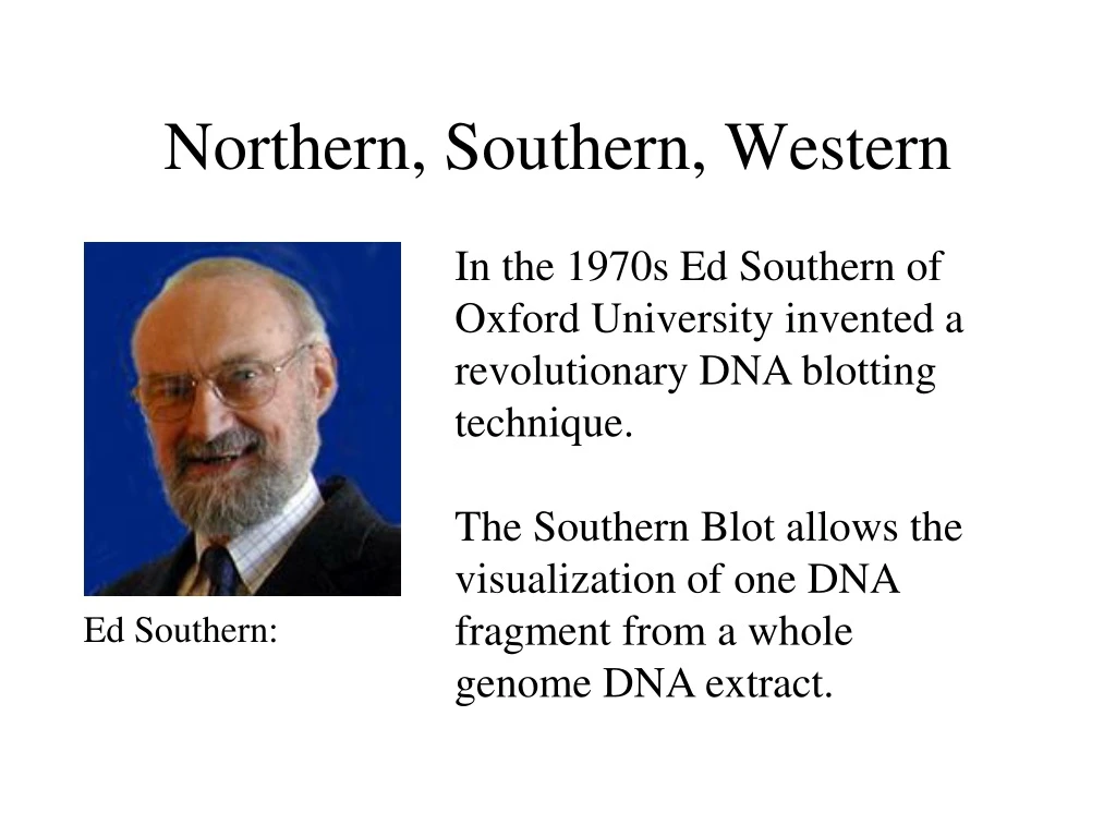
Northern, Southern, Western. In the 1970s Ed Southern of Oxford University invented a revolutionary DNA blotting technique. The Southern Blot allows the visualization of one DNA fragment from a whole genome DNA extract. Ed Southern:. Northern and Western.

E N D
Northern, Southern, Western In the 1970s Ed Southern of Oxford University invented a revolutionary DNA blotting technique. The Southern Blot allows the visualization of one DNA fragment from a whole genome DNA extract. Ed Southern:
Northern and Western • People then applied the same technique to RNA. • They called it a “Northern blot”. • Then other people applied it to protein, and imaginatively called it a “Western blot” Funny, eh?
Introduction Concept: reannealing nucleic acids to identify sequence of interest. Separates DNA/RNA in an agarose gel, then detects specific bands using probe and hybridization. Hybridization takes advantage of the ability of a single stranded DNA or RNA molecule to find its complement, even in the presence of large amounts of unrelated DNA. Allows detection of specific bands (DNA fragments or RNA molecules) that have complementary sequence to the probe. Size bands and quantify abundance of molecule.
Southern Blot: DNA-DNA* Developed by Edwin Southern. Uses gel electrophoresis together with hybridization probes to characterize restriction fragments of genomic DNA (or DNA from other sources, such as plasmids). Identifies DNA with a specific base sequence. Can be done to detect specific genes present in cells.
Southern Steps 1. DNA to be analyzed is digested to completion with a restriction endonuclease. 2. Electrophoresis to maximally separate restriction fragments in the expected size range. A set of standards of known size is run in one lane of the gel. 3. Blot fragments onto a nitrocellulose membrane. 4. Hybridize with the 32P probe. 5. Autoradiography.
Step 2 Gel electrophoresis • Separates DNA fragments. Soak gel in 0.5 M NaOH • Converts dsDNA to ssDNA
Step 3. Nitrocellulose Blot • Cover gel with nitrocellulose paper…then… • Cover nitrocellulose paper with thick layer of paper towels. • Compress apparatus with heavy weight. • ssDNA binds to nitrocellulose at same position it had on the gel. • Vacum dry nitrocellulose at 80C to permanently fix DNA in place or cross link (via covalent bonds) the DNA to the membrane.
Step 4. Hybridization • Incubate nitrocellulose sheet with a minimal quantity of solution containing 32P-labeled ssDNA probe. • Probe sequence is complementary to the DNA of interest. • Incubate for several hours at suitable renaturation temperature that will permit probe to anneal to its target sequence(s). • Wash & dry nitrocellulose sheet.
Step 5. Autoradiography • Place nitrocellulose sheet over X-ray film. • X-ray film darkens where the fragments are complementary to the radioactive probes.
Characterization: Southern blot hybridization -transfer of DNA from a gel to a membrane (e.g., nitrocellulose, nylon) -developed by Edwin Southern
Southern Application: Diagnosis & detection of genetic diseases. • Used to diagnose sickle cell-anemia. • AT base change in the subunit of Hb Glu Val. • Development of a 19 residue oligonucleotide probe complementary to sickle-cell gene’s mutated segment. • Probe hybridizes to DNA from homozygotes of sickle-cell anemia but not from normal individuals.
Northern Blot: RNA-DNA*(RNA*) • Alwine adapted Southern's method for DNA to detect, size and quantify RNA – 1977. • No need to digest RNA with restriction enzymes. • Use formaldehyde to break H-bonds and denature RNA because single-stranded RNA will form intramolecular base pairs and "fold" on itself.
Northern Steps 1. Isolate RNA & treat with formaldehyde. 2. Electrophorese RNA in denaturing agarose gel (has formaldehyde). Visualize RNA in gel using Ethidiumbromide stain and photograph. 3. Transfer single-stranded RNA to nitrocellulose or nylon membrane. Covalently link RNA to membrane. 4. Incubate membrane (RNA immobilized on membrane) with labeled DNA or RNA probe with target sequence. 5. Development.
Step 1 Isolate RNA: -To detect rare mRNA, isolate the poly A+ mRNA. -RNA is both biologically and chemically more labile than DNA. Thus eliminate RNases. Step 2 Electrophoresis: - Performed in formaldehyde agarose gel to prevent RNA from folding on itself. - Stain with EtBr to visualize the RNA bands.
Step 3 -Transfer single-stranded RNA to nitrocellulose or nylon membrane: Traditionally, a nitrocellulose membrane is used, although nylon or a positively charged nylon membrane may be used. Nitrocellulose typically has a binding capacity of about 100µg/cm, while nylon has a binding capacity of about 500 µg/cm. Many scientists feel nylon is better since it binds more and is less fragile. -Covalently link RNA to membrane: UV cross linking is more effective in binding RNA to the membrane than baking at 80C.
Step 4 & 5 -Prehybridize before hybridization: Blocks non-specific sites to prevent the single-stranded probe from binding just anywhere on the membrane. -Incubate membrane with labeled DNA or RNA probe with target sequence: Probe could be 32P, biotin/streptavidin or a bioluminescent probe. -Autoradiography: Place membrane over X-ray film. X-ray film darkens where the fragments are complementary to the radioactive probes.
Northern Application • Northern blots are particularly useful for determining the conditions under which specific genes are being expressed, including which tissues in a complex organism express which of its genes at the mRNA level. • For instance: When trying to learn about the function of a certain protein, it is sometimes useful to purify mRNA from many different tissues or cell types and then prepare a Northern blot of those mRNAs, using a cDNA clone of the protein of interest as the probe. Only mRNA from the cell types that are synthesizing the protein will hybridize to the probe.
Southern DNA on membrane. Digest DNA. Convert dsDNA to ssDNA. Probe with DNA or RNA. Northern RNA on membrane. No need to digest DNA. Denature “folded” RNA with formaldehyde. Probe with DNA or RNA. Summary
Characterization: Western blotting X Protein Enzyme reaction or X React with Antibody X x Buffer; requires electric current X -transfer of protein from a gel to a membrane (e.g., nitrocellulose, nylon) -requires the use of an electric current to facilitate transfer
Fluorescence in situ Hybridization (FISH) • FISH - a process which vividly paints chromosomes or portions of chromosomes with fluorescent molecules. Identifies chromosomal abnormalities • Aids in gene mapping, toxicological studies, analysis of chromosome structural aberrations, and ploidy determination
FISH • Used to identify the presence and location of a region of DNA or RNA within morphologically preserved chromosome preparations, fixed cells or tissue sections. • This means you can view a segment or entire chromosome with your own eyes • Was often used during M phase but is now used on I phase chromosomes as well
FISH • Advantage: less labor-intensive method for confirming the presence of a DNA segment within an entire genome than other conventional methods like Southern blotting.
FISH Procedure • Denature the chromosomes • Denature the probe • Hybridization • Fluorescence staining • Examine slides or store in the dark.
Probes Fluorescein Complementary sequences of target nucleic acids • Designed against the sequence of interest • Probes are tagged with fluorescent dyes like biotin, fluorescein, Digoxigenin • Size ranges from 20-40 bp to 1000bp Biotin
FISH Uses • Detection of high concentrations of base pairs • Eg: Mouse metaphase preparation stained with DAPI (4',6-diamidino-2-phenylindole) is a fluorescent stain that binds strongly to A-T rich regions in DNA. It is used extensively in fluorescence microscopy.
FISH Uses Centromere regions stained brighter - means they are rich in A-T bonds Also used in germ cell or prenatal diagnosis of conditions such as aneuploidies (abnormal number of chromosomes in a cell).
FISH and Telomeres • Telomeric probes define the terminal boundaries of chromosomes (5’ and 3’ ends) • Used in research of chromosomal rearrangements and deletions related to cell aging or other genetic abnormalities. • Special telomeric probes specific to individual chromosomes have been designed. • Probe is based on the TTAGGG repeat present on all human telomeres.
FISH and Telomeres Application in cytogenetics - can detect submicroscopic deletions and cryptic translocations of genes associated withunexplained mental retardation and miscarriages
FISH - Medical FISH can be used in the study of transgenic animals (eg: Polly) Selective markers show if the human DNA was inserted successfully and pinpoint where the human DNA is .
Diagnostic Applications of FISH • Prenatal diagnosis • Cancer diagnosis • Molecular cytogenetic of birth defects and mental • retardation • The identification of specific chromosome • abnormalities • • The characterization of marker chromosomes • • Interphase FISH for specific abnormalities in cases of • failed • • Cytogenetic • • Monitoring the success of bone marrow transplantation
Other Human Disorders • Sickle Cell Anemia – • autosomal recessive • Found most often among people of African ancestry • Blood cells sickle (change shape) when oxygen-deprived (exertion, increase in altitude) • Causes sickle cell event – pain and immobility and death of tissue ( dangerous if in organ) • Treatment – hospitalization and oxygen • Carriers are resistant to malaria
Tay Sachs Disease • Autosomal recessive ( Gangliosidosis or Hexosaminidase A deficiency) . • Genetic mutation of HEXA gene on chromosome 15 • Found most often among Jews of Mediterranean ancestry • Child born appearing normal, but fat builds up in brain and child dies by age 5 • No treatment, no cure
Huntington Disease • It is a neurodegenerative genetic disorder that affects muscle coordination and leads to cognitive decline and dementia • Autosomal dominant • Symptoms do not appear until age 30-40. • Death takes about 5-10 years • No treatment, no cure – but there is a test to see if you have it before symptoms begin • Results in mental impairment and uncontrollable spastic movements
Deletions • Chromosome fragment breaks off and is lost • Cri du chat syndrome – mental retardation and cat-like cry, delayed development, distinctive facial features, small head size (microcephaly), widely-spaced eyes (hypertelorism), low birth weight and weak muscle tone (hypotonia) in infancy. • It is a rare genetic condition that is caused by the deletion (a missing piece) of genetic material on the small arm (the p arm) of chromosome 5.
The condition affects an estimated 1 in 20,000 to 50,000 live births, strikes all ethnicities, and is more common in females by a 4:3 ratio
Prenatal Tests to detect chromosomal problems: • Amniocentesis – removes a little amniotic fluid from around baby – fluid is then tested for abnormal proteins and the cell in it can be karyotyped. • Amniocentesis usually done between 16 to 20 weeks, which is during second trimester. • Risk of miscarriage
Chorionic Villus Sampling • Take a piece of the chorionic villus from the placenta – it is made of baby cells – and test as in amniocentesis It is done during early pregnancy, most often between the 10th and 12th weeks. • Risk of miscarriage • Has been linked to deformed fingers
Bioethical Dilemma • Once a prenatal diagnosis of a genetic disorder is made, what are the parents going to do? • Do nothing and give birth to child with disorder • Abort embryo/fetus • Who should make the decision? • What should enter into making the decision?
Genetic Counseling • Genetic counselor: • educates the parents about the disorder, • tells them of their options without influencing their decision, • and tells them of the consequences of each option .
DNA Fingerprints • The pattern of DNA formed during gel electrophoresis. Used by law enforcement.
“DNA in the Courtroom” 1. Use of VNTRs (variable number of tandem repeats; different individuals have different numbers of repetitive stretches of DNA, for example, GGAGG). One individual might have 6, another 12. 2. VNTRs can be analyzed by gel electrophoresis, creating a banding pattern specific to each individual—like a bar code .
END Part II