Download
1 / 24
280 likes | 569 Views
Department of Neutron Activation Analysis Division of Nuclear Physics Frank Laboratory of Neutron Physics Joint Institute for Nuclear Research. Biosynthesis of silver and gold nanoparticles using microbial biomass. Inga Zinicovscaia E-mail: zing@nf.jinr.ru.

E N D
Department of Neutron Activation Analysis Division of Nuclear Physics Frank Laboratory of Neutron Physics Joint Institute for Nuclear Research Biosynthesis of silver and gold nanoparticles using microbial biomass Inga Zinicovscaia E-mail: zing@nf.jinr.ru JINR Scientific Council, September 15-16, 2011
M.V. Frontasyeva, S.S. Pavlov • Frank Laboratory of Neutron Physics , • JINR, Russian Federation • T.Kalabegishvili , E. Kirkesali,I. Murusidze , • D. Pataraya, E.N. Ginturi • Andronikashvili Institute of Physics, Tbilisi , Georgia • I. Zinicovscaia, Gh. Duca Institute of Chemistry of the Academy of Science of Moldova, Chisinau, Moldova
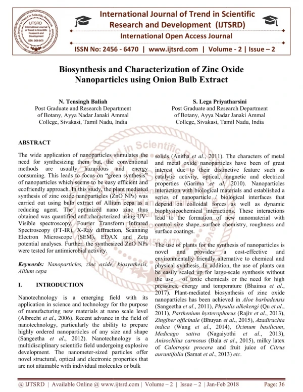
Introduction An important area of research in nanotechnology deals with synthesis of nanoparticles of different chemical composition and size. There is a growing need to develop environmentally gentle nanoparticle synthesis that does not use toxic chemicals in its process. As a result, researchers in the field of nanoparticle synthesis have turned to biological systems. It is well known that many organisms, both uni-cellular and multi-cellular, are producing inorganic materials either intra- or extra-cellularly. 3
Advantages of biological method • tightly controlled, highly reproducible syntheses • biocompatible particles • the avoidance of toxic surfactants or organic solvents 4
Application of silver nanoparticles • nonlinear optics • medicine • electronics • catalysis • microelectronics 6
Experiment = AgNO3 1mM + yellowish brown pale yellow UV-Vis spectra recorded after one week for the reaction mixture prepared using 1mM silver nitrate and 1 g Streptomyces glaucus 71MD 7
Scanning electron microscope Resolution 1.2 nm Magnification 5000–150000x Voltage 1–30 kV Quanta 3D FEG The Netherlands’ Firm “Systems for Microscopy and Analysis” (Moscow, Russia) 8
Objects of study p Bacteria Streptomyces glaucus 71MD Blue-green microalga Spirulina platensis 9
1 day Control 27 nm SEM micrographs of Spirulina platensis cells with silver nanoparticles 10
C O EDAX spectrum recorded from Spirulina platensis cells after formation of silver nanoparticles 11
150 000x 50 000x Control 12 000x SEM micrographs of Streptomyces glaucus 71MD cells with silver nanoparticles 12
EDAX spectrum of Streptomyces glaucus 71MD cells after exposure to silver nitrate solution 13
Biotechnology of • gold nanoparticles
Application of gold nanoparticles • Catalysis • Chemical sensing • Biosensing • Medicine 15
Experiment HAuCl4 + = red purple yellow Experiment I CHAuCl4= 1mM Incubation time: 1 − 6d Experiment II Incubation time = constant CHAuCl4: 10-4 − 10-2M 16
UV-Vis spectra recorded after one week for the reaction mixture prepared using 1mM hydrated gold chloride and 1 g biomass of A. globiformis 151B 17
Experiment I 2 days 3 days 1 day Control 5 days 6 days SEM micrographs of Spirulina platensis cells with gold nanoparticles at different incubation time 18
Experiment II 10-2M HAuCl4 10-3M 10-4M 5·10-3M SEM micrographs of Spirulina platensis cells with gold nanoparticles at different concentrations 19
EDAX spectrum of Sp. platensis cells after exposure to hydrated gold chloride solution 20
Conclusions 1. Production of silver and gold nanoparticles by blue-green microalgae Spirulina platensis and bacteria Streptomyces glaucus 71MD proceeds extra-cellularly 2. SEM and EDAX were used to characterize the silver and gold nanoparticles. SEM showed formation of nanoparticles in the range of: 4–25 nm for Streptomyces glaucus 71MD 16–200 nm for Spirulina platensis (first experiment) 15 nm–7 μm for Spirulina platensis (second experiment) 21
For quantitative analysis of samples the epithermal neutron activation analysis (ENAA) in the radioanalytical complex REGATA at the reactor IBR-2M will be carried out by the end of 2011 22
Elemental concentration in biomass of Streptomyces glaucus (irradiation time 8 s) The data were obtained by Sergey Pavlov (FLNP JINR) and Arnaud Faanhof (NECSA) in June 2011 at the reactor SAFARI-I 23