Download
1 / 12
120 likes | 274 Views
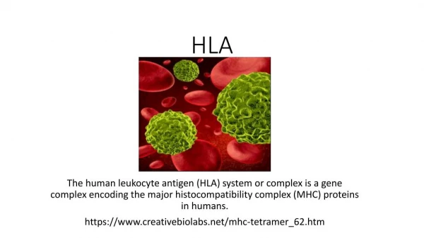
Assay I HLA-DQ Alpha Haplotype. Purpose. To determine which one of several known alleles is present at the HLA DQ-Alpha locus on each of an individual’s two chromosomes Why? Tissue typing Identifying genetic relatives Suspect identification Is this assay qualitative or quantitative?.

E N D
Purpose • To determine which one of several known alleles is present at the HLA DQ-Alpha locus on each of an individual’s two chromosomes • Why? • Tissue typing • Identifying genetic relatives • Suspect identification • Is this assay qualitative or quantitative?
Sample • Start with human hair follicle cells • Any human cells containing nuclear DNA • PCR the HLA DQ-Alpha region • PCR is an enzymatic technique used to increase the amount of sample
Detection Scheme • Probes - 8 oligonucleotides each with different sequence • Affixed to membrane strip • Sequence of each probe is that of a different HLA DQ-alpha allele
Detection Scheme • Specificity - complementary base pairing • Precise hybridization conditions limit pairing to exact complements • Hybridization = complementary base pairing of two single DNA or RNA strands to each other • PCR products from 1 person’s sample base pair with only 1 or 2 probes • 1 - homozygous • 2 - heterozygous
Visualization • Color reaction visible to naked eye • Color created by enzyme/substrate reaction • Enzyme = Horseradish peroxidase • Chromogen Substrate = TMB (tetramethylbenzidine) • Reaction = 2H2O2 + TMB (colorless) 3H2O + oxidized TMB (blue)
How is the color localized to the Probe/PCR product complex? • PCR product includes biotin • Primers are biotinylated • Enzyme is conjugated to streptavidin • Conjugated = covalently bound • Streptavidin binds to biotin • So . . . enzyme/streptavidin conjugate localizes where PCR product is base-paired to probe • And . . .colored product is made and precipitates only where enzyme is
Contributions to sensitivity • Sample amplification by PCR • Signal amplification by repetitive enzymatic catalysis to yield more and more color • Background reduction • Blocking - prevents non-specific interactions • Rinsing - removes excess and misbound reagents • Note: Blocking and rinsing also contribute to specificity
Controls • PCR reaction • Positive control template – controls for failure of PCR reagents • Template negative – controls for presence of contaminant DNA • Agarose gel electrophoresis – checks for PCR yield • Hybridization • Control PCR product • Checks for hybridization success and specificity • Control probe on each strip • Warns of potential for false interpretation
Ask yourself • How has each of the essential assay components been incorporated into this assay?