Download
1 / 25
250 likes | 393 Views
Potential Control Scenarios. Tracy Rausch, CCE. Purpose. Select a set of Clinical Scenarios to point to derive a set of control Use Cases to Study Add as an annex to Remote Control Standard. Clinical Scenarios. PCA Safety Interlock OR to ICU Room Readiness

E N D
Potential Control Scenarios Tracy Rausch, CCE
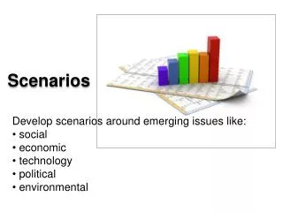
Purpose • Select a set of Clinical Scenarios to point to derive a set of control Use Cases to Study • Add as an annex to Remote Control Standard
Clinical Scenarios • PCA Safety Interlock • OR to ICU Room Readiness • Imaging vs Ventilator Synchronization (OR and ICU) • Remote Control for Isolation Room • Closed Loop Control During Procedural Sedation • Glucose Management (Inpatient, and Home) • Medication Management (Heparin) • Airway Fire Prevention
PCA Safety Interlock • Video Was here see www.mdpnp.org website
Proposed State • Video Was here see www.mdpnp.org website
OR to ICU Handoff – Room Readiness • Video Was here see www.mdpnp.org website
Proposed State • Video Was here see www.mdpnp.org website
Example: Cholecystectomy (gall bladder removal) w/ intraop cholangiography (x-ray) Workflow: 1) Ventilation is stopped. 2) Intraoperative cholangeogram is performed with contrast to identify internal structures. Breath hold -> improve x-ray quality. Ventilator X-ray
“With the advent of sophisticated anesthesia machines incorporating comprehensive monitoring, it is easy to forget that serious anesthesia mishaps still can and do occur.” APSF Newsletter Winter 2005 A 32-year-old woman had a laparoscopic cholecystectomy performed under general anesthesia. At the surgeon’s request, a plane film x-ray was shot during a cholangiogram. The anesthesiologist stopped the ventilator for the film. The x-ray technician was unable to remove the film because of its position beneath the table. The anesthesiologist attempted to help her, but found it difficult because the gears on the table had jammed. Finally, the x-ray was removed, and the surgical procedure recommenced. At some point, the anesthesiologist glanced at the EKG and noticed severe bradycardia. He realized he had never restarted the ventilator. This patient ultimately expired.
Synchronize x-ray with ventilator:@ expiration: cholangiogram, angiograms@inspiration: routine chest radiograph In this case, integration of devices into a networked, smarter system can improve safety by avoiding ventilator shut-off, improve image quality (especially on serial images), and decrease re-imaging.
Interventional Radiology I better get out of here!
Remote Control of Isolation/Burn Unit A patient is in isolation which requires clinicans to use precautions when caring for the patient. A remote panel is located outside the room and the clinician can change settings and silence alarms on devices from outside the room.
GI Procedural Sedation • Autonomous control: use SpO2, ETCO2, BIS data for: • Safety interlock – e.g. Stop IV propofol pump if resp or BP low, or • Physiologic Closed Loop Control - Titrate infusion rate of IV propofol pump to target BIS value • And, create smart alarm and activate innovative alarm signal
Glucose Management • Utilizing Physiological Parameters and Real-Time POC Glucose to manage rate of insulin infusion.
Inpatient Glucose A patient is receiving IV insulin via a syringe pump, glucose solution via a large-volume infusion pump, and a large-volume infusion pump of saline serving as the carrier line. The patient is also attached to a continuous blood glucose monitor or an intermittent glucose monitor. At the time of connecting the patient to an IV infusion, the nursing staff completes assessments of vital signs and IV line integrity. Subsequently, the large volume infusion pump (saline carrier), syringe pump (insulin), and blood glucose monitor are attached to an integrated system that queries the patient record for weight, target glucose range, typical insulin dosage range (and correction factor), and glucose responsiveness to meals (insulin-to-carbohydrate ratio). The integrated system hosted PCLC algorithm delivers IV insulin to maintain the blood glucose values within the clinically desired range. The clinical staff is alerted if the glucose level changes unexpectedly or outside the limits determined by the system. In order to maintain the glucose levels within the target range, the system can 991 also change the glucose infusion rate utilizing an integrated system-hosted algorithm. The algorithm would alert the clinical staff if the glucose levels exceed a range that the algorithm can effectively manage by adjusting the insulin or glucose infusions.
Medication Management The patient is attached to a large volume infusion pump with heparin solution. During the setup of the large volume infusion pump, the dosage of the heparin IV bag is verified with the computerized provider order entry system. Heart rate, blood pressure, and respiration rate are measured. An IV line assessment is completed. When the integrated system recognizes that the medication being infused is heparin, it automatically places an order for serial PTT tests. Once the laboratory information system determines the PTT, the integrated system retrieves the results and an integrated system-hosted algorithm determines whether changes to the dosage need to be made, and the clinical staff is notified.
Scenario: Surgical Fires 600 surgical fires each year The most severe burns are internal – in the lungs Caused by burning breathing tubes
Airway Laser Surgery + O2 -> Fire • O2 in respiratory gas supports combustion. • If laser hits breathing tube, could produce devastating burn. • Surgical team must “remember” to minimize O2 Breathing Tube with Oxygen
A Solution: Laser-O2 Interlock or smart alarm • Monitor ventilator O2 concentration. (This is already measured.) • “Smart” safety interlock to prevent laser activation if O2 > 25%
A Solution: Laser-O2 Interlock or smart alarm • Monitor ventilator O2 concentration. (This is already measured.) • “Smart” safety interlock to prevent laser activation if O2 > 25%
Clinical Scenarios • PCA Safety Interlock • OR to ICU Room Readiness • Imaging vs Ventilator Synchronization (OR and ICU) • Remote Control for Isolation Room • Closed Loop Control During GI procedure • Closed Loop Control of Anesthetic • Glucose Management (Inpatient, and Home) • Medication Management (Heparin) • Airway Fire Prevention
Control High Level Use Cases • Transfer/Change of Device Settings During Configuration State • Safety Interlock – Remotely changing the state of a device • Transfer/Change of Device Setting During Operating State • Physiological Closed Loop Control