Download
1 / 44
440 likes | 460 Views
HEARING CONSERVATION PROGRAM. OTOSCOPIC EXAMINATION and TYPANOMETRY BASICS. 28 Jan 2013. Learning Objectives. Explain the purpose of otoscopic examination and tympanometry Describe the basic characteristics of a normal and an abnormal tympanogram

E N D
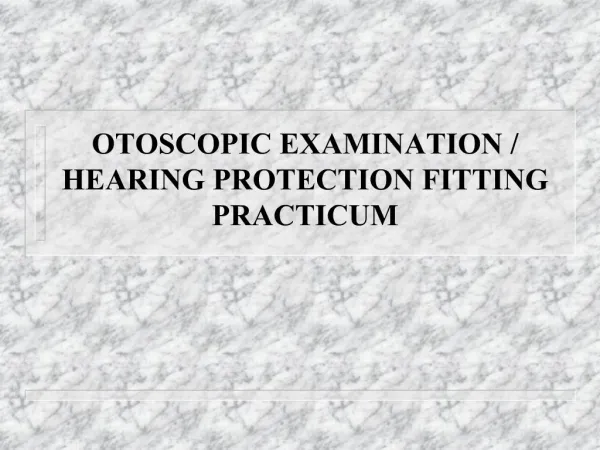
HEARING CONSERVATION PROGRAM OTOSCOPIC EXAMINATIONandTYPANOMETRY BASICS 28 Jan 2013
Learning Objectives • Explain the purpose of otoscopic examination and tympanometry • Describe the basic characteristics of a normal and an abnormal tympanogram • Describe normal and abnormal physical findings • State conditions requiring medical referral • Perform otoscopy and tympanometry using proper technique
OTOSCOPIC EXAMINATION Definition The examination of the ear canal and tympanic membrane through the use of an otoscope. An otoscope is a hand-held tool with a speculum and light source to see into the ear canal Purpose The otoscopic exam is to ensure that the ear canals are free of any obvious problems prior to fitting hearing protection, performing tympanometry and administering hearing tests
Otoscope Check • Check if batteries are fully charged • Adjust rheostat to bright white light • Fiber-optic better than older bulb-types
Preparation for Otoscopic Exam 1. Observe proper hygiene wash hands or use gloves note any bodily fluid or secretion 2. Select a speculum of proper size larger size ensures a good view 3. Lock speculum into place 4. Change/discard the speculum after each patient after each ear of any patient with draining ear(s)
Examination Method KEY: #1 Otoscope placement #2 Eye placement • Grip otoscopefirmly and comfortably • Grasp upper edge of the ear (helix)with the opposite hand • Pull pinna gently upward & back to straighten the ear canal • Insert lighted otoscope past the first canal bend • Rest your fingers against the patient’s head to avoid injury if patient moves suddenly • NOW put your eye up to the otoscopeeyepiece
Examination Method cont… • Examine the ENTIRE canal and tympanic membrane • Dispose speculum, turn off otoscope light Don’t be satisfied with a partial viewing NO discomfort to the patient if properly conducted YOUR GOAL “Within Normal Limits” or “Abnormal” Do not diagnose or label pathology
What is Tympanometry? DEFINITION A measurement technique that assesses function of the middle ear The technique uses • an acoustic input signal • air pressure • electronic measurement
Tympanometry Rules OutDisorders of the Middle Ear Outer Ear Middle Ear
Why use Tympanometry? PURPOSE • To identify patients who require medicalreferral for middle ear pathology • To differentiate conductive from sensorineural hearing disorders • To track the progress of middle ear pathologies under medical treatment • Fast, objective, highly accurate
When will you use Tympanometry? PROTOCOL • After otoscopy • Part of the referral procedure • If positive STS is present • If patient complains of ear fullnessor pressure NEVER USE TYMPANOMETRY WHEN THERE HAS BEEN MIDDLE EAR (BONE) SURGERY
How Does Tympanometry Work? • Probe inserted and seals ear canal • Pump varies pressure against eardrum • Pure tone is sent into ear • Tympanometer measures how much sound gets through the eardrum • Results indicate the flexibility of the eardrum and middle ear
Normal Tympanogram“Type A” Inverted “V” or Mountain Peak placement Horizontal Vertical -150 to +50 0.2 to 1.8 Display uses “box” or shaded area to show normal range
Interpretation – Type A Normal eardrum movement Normal middle ear pressure Eustachian Tube is functioning normally Normal Outer/Middle Ear No conductive HL STS is inner ear related
Variations of Normal Type A Peak Horizontal -150 to +50 Vertical 0.2 to 1.8
Abnormal Tympanogram“Type B” Flat or poorly defined peak Peak Placement Absent or poorly defined Horizontal Vertical > -150 < 0.2
Interpretation - Type B Eardrum movement minimal or absent Outer and/or middle ear disorder present Ear canal may be occluded Eustachian Tube not functioning normally Otitis Media or ME Effusion Eardrum has perforation Mild hearing loss in low Hz
Abnormal Tympanogram“Type C” Inverted “V” off center to left Clearly defined peak Peak placement Horizontal Vertical > -150 0.2 to 1.8
Interpretation - Type C Negative middle ear pressure Eustachian Tube function abnormal Recent air flight or diving Symptoms of congestion Hearing normal or slight loss @ 500-1000Hz
Disposition of Patients with Abnormal Tympanograms General Rule Medical referral Final Decision Local resources and SOP To Determine Referral Request return for follow-up tympanogram Otitis Media or Middle Ear Effusion onset to resolution Tympanograms can progress Type C >> Type B >> Type C >> Type A over 10-14 day period
Referral Protocol Refer to MO or Audiology Abnormal Action required Test again 10-14 days Normal No action required
Referral to Medical Officer when... SUMMARY • Pain or discomfort is reported • Drainage is visible • Perforation is visible • Tympanic membrane is bulging • Ear canal is blocked by cerumen or foreign body • Complaint of sudden severe hearing loss with tinnitusand/or dizziness • When in doubt STAT!
OHC Technician Responsibilities SUMMARY • Always perform otoscopy first • Interpret tympanograms as “Normal” or “Abnormal” only • Consider all information before referral • Patient history • Otoscopy • Tympanogram • Audiograms
“Within Normal Limits” Ear Canals clear and free of obvious problems discharge, masses, impacted cerumen, foreign bodies, inflammation Tympanic membrane appearance translucent, pearly gray healthy color Eardrum landmarks Cone of light from center to membrane edge Shadow of first middle ear bone attached to center Cerumen is normal unless occludes view of TM > 50%
“Within Normal Limits” Type A Normal Peak Normal Pressure
Excessive Cerumen If you can’t see at least half the TM, then cleaning is recommended If the TM is normal, proceed with hearing test and refer for removal Type A If not fully occluded Type B If fully occluded Photo’s courtesy of Dr. Roy F. Sullivan, Ph.D.
Foreign Bodies Insect on Canal Wall Shattered Glass Type A Normal Peak Normal Pressure unless foreign body fully occludes canal Photo’s courtesy of Dr. Roy F. Sullivan, Ph.D.
Cotton Swab/Earplug Residue Earplug One Year After Rock Concert Cotton Swab Residue Type A Normal Peak Normal Pressure unless foreign body fully occludes canal Photo’s courtesy of Dr. Roy F. Sullivan, Ph.D.
Collapsing Canals When patients display a “flat” hearing loss, rule out by observing ear canal as you press pinna Type A Normal Peak Normal Pressure tension of headphone collapses canal Photo’s courtesy of Dr. Roy F. Sullivan, Ph.D.
Exostoses Will not affect the hearing test unless ear canal fully occluded Interferes with earplug insertion Interferes with otoscopy Type A Normal Peak Normal Pressure unless fully occludes canal Photo’s courtesy of Dr. Roy F. Sullivan, Ph.D.
Eardrum Perforations Type B Flat Large Ear Canal Volume Size of hole will affect hearing test
Retracted Eardrum Type C Abnormal Negative Pressure Photo’s courtesy of Dr. Roy F. Sullivan, Ph.D.
OtitisMedia / Middle Ear Effusion Type B Flat - no peak Normal ear canal volume Photo’s courtesy of Dr. Roy F. Sullivan, Ph.D.
Pressure Equalization (PE) Tubes or Ventilation Tubes Type C Flat straight line Huge ear canal volume Photo’s courtesy of Dr. Roy F. Sullivan, Ph.D.
Other Middle Ear Diseases • Tymp Types Vary • depends on • stiffness of TM • & • size of mass • in middle ear Cholesteatoma Tympanosclerosis
Patient Scenario #1 • Patient Complaints : congestion recent head cold • Postive STS • Large threshold shifts at 500Hz & 1000Hz • Otoscopy – slightly red TM Do you refer?
Patient Scenario #2 • Patient Complaints: sounds are muffled gradually noticed it • Postive STS for most frequencies • Otoscopy - cerumen in canal --unsure if fully occluded Do you refer?
Patient Scenario #3 • Patient Complaints: feels plugged up recent airplane flight • Positive STS, decreased hearing in lower frequencies. • Otoscopy eardrum good color ossicles very visible Do you refer?
Patient Scenario #4 • Patient Complaints: trouble hearing in noise ringing in both ears • Positive STS in high frequencies • Otoscopy clear canal TM normal color Do you refer?
Otoscopyand Tympanometry SUMMARY • Otoscopic examination reveals condition ear canal and eardrum • Tympanometry is a fast, objective, accurate assessment of middle ear status and function • A Tympanometer measures eardrum movement during controlled pressure changes • A Tympanogram shows results in graphic form • Refer to medical officer or audiologist for possible conductive disorders