Download

1 / 69
690 likes | 698 Views
MUSCLE PHYSIOLOGY REVIEW. 1. – 65. 1. Name this protein. actin. 2. Name this specific band. H Band. 3. Name this unit. Fascicle (fasciculus). 4. Name this unit. Myofiber (muscle cell). 5. Name this protein. myosin. 6. Name bluish CT layer. perimysium. 7. Name this dark line.

E N D
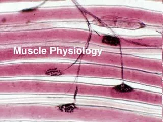
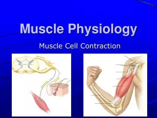
MUSCLE PHYSIOLOGY REVIEW 1. – 65.
1. Name this protein. actin
2. Name this specific band. H Band
3. Name this unit. Fascicle (fasciculus)
4. Name this unit. Myofiber (muscle cell)
5. Name this protein. myosin
6. Name bluish CT layer. perimysium
7. Name this dark line. M line
9. Name these purple structures. mitochondria
10. Name this entire structure. myofiber
11. Name these blue structures. Sarcoplasmic reticulum
12. Name these yellow structures. Transverse tubules
13. Name this unit. triad
14. Name this entire unit; it is the smallest unit of a muscle contraction (red bracket). sarcomere
15. Name the cytoplasm inside sarcoplasm
16. Name this covering. muscle epimysium
2 3 17. Which is the crossbridge? 1 4 4
1 2 18. Which is in the contacted state? 2
21. What is the SPECIFIC role of this molecule in muscle contraction? Allows myosin to perform a(n)__________ so actin is pulled inward. power stroke
22. What is the name of the 3 purple protrin complex? troponin
23. What happens to the width of the A band during contraction? Stays the same
What condition are these muscles in? hypertrophy
25. What is represented by the green circles in this diagram? calcium
26. Where specifically is calcium stored in the muscle? Sarcoplasmic reticulum
27. What is the name of the gray molecule? tropomyosin
28. What is the other source for recharging the ATP battery in the muscles? (this compound is unique to muscles) Creatine phosphate
29.What is the specific function of calcium in a muscle contraction? Calcium ions bind to_________ which causes a change in the conformation of the tropomyosin complex that exposes the myosin binding sites on the actin filament. troponin.
30. What is this known as? Sliding filament theory
31. What is this set-up called? Motor unit
32. The products of aerobic respiration are water, ATP, and _____. Carbon dioxide
33. Name this red area shown by red arrow. Motor end plate
34. Name this yellow structure of which you see the end of. Motor neuron
35. Name these blue “containers”. Synaptic vescicles
36. What is the general name for the compound that is in these blue “containers”. neurotransmitter
37. Name these structures shown by red arrows. Transverse tubules
38. Name this entire area shown by bracket. Neuromuscular junction
39. During the contraction of a sarcomere, calcium ions bind with the protein _____. troponin
40. This is a graph of a muscle contraction. What is it called? myogram
41. Name this blue part of a muscle twitch. Latent period
42. Name this red part of a muscle twitch. Contraction period Contraction period
43. What is the bracketed part of this graph representing dealing with a muscle contraction? Treppe or summation
Tetanus (tetanic contraction) 44. What is the bracketed part of this graph representing dealing with a muscle contraction?
45. Choose the type of muscular contraction shown below. isotonic
46. The reddish brown color of muscle is due to the presence of _____ molecules myoglobin
47. Which molecule is produced during exercise, resulting in the oxygen debt? Lactic acid
48. Would there be mostly slow twitch or fast twitch fibers in this part of your Thanksgiving Turkey? Fast twitch
49. The minimum stimulus needed to cause a contraction is called the _____. Threshold stimulus