Download

1 / 27
270 likes | 458 Views
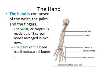
The Hand. Sophie Taylor. Anatomy. Radius. Ulnar. Carpals. Metacarpals. Proximal, Intermediate, Distal Phalanges. S ome L overs T ry P ositions T hat T hey C an’t H andle. S caphoid L unate T riquetral P isiform T rapezium T rapezoid C apitate H amate.

E N D
The Hand Sophie Taylor
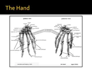
Radius Ulnar Carpals Metacarpals Proximal, Intermediate, Distal Phalanges
Some Lovers Try Positions That They Can’t Handle Scaphoid Lunate Triquetral Pisiform Trapezium Trapezoid Capitate Hamate
Abductor Pollicis Longus Extensor Pollicis Brevis Dorsal and Palmer Interossei
Abductor Pollicis Longus Extensor Pollicis Brevis Adductor Pollicis: Oblique Head Adductor Pollicis: Transverse Head
Flexor DigitorumProfundus & Superficialis Superficialis Flexor Pollicis Longus Profundus
Extensor PolicisLongus Extensor Digitorum Extensor Policis Brevis Extensor DigitiMinimi Extensor Indicis
Flexor DigitiMinimi Brevis OpponensDigitiMinimi Abductor DigitiMinimi
Abductor Pollicis Brevis Opponens Pollicis Flexor Pollicis Brevis The 2 Lateral Lumbricals LOAF Muscles
Radial Artery Ulnar Artery Median Artery Superficial Palmar Arch Dorsal Carpal Arch Metacarpal Arteries Palmar Digital Arteries Arterial Supply
Median Vein Cephalic Vein Basilic Vein Palmer and Dorsal Venous Plexii Palmer and Dorsal Digital Veins
Nerves Pathways, Functions, & Palsies
Nerve Root Impingement • Pain • Paraesthesia • Numbness In dermatome • Weakness • Wasting • Fasciculation In muscles supplied by that segment May also complain of neck pain in the corresponding segment, or have suffered a trauma to the neck
Ulnar Nerve • Nerve Roots: C7, C8, T1 • Muscles Supplied: • Interossei • Medial Lumbricals • Hypothenar Eminence • Flexor Carpi Ulnaris • Medial half of Flexor DigitorumProfundus • Places of impingement: • First Rib/Cervical Rib • Pectoralis Minor • Ulnar Groove, medial elbow • Elbow dislocation • Two heads of Flexor Carpi Ulnaris • Tunnel of Guyon • Palsy: • Muscle Wasting, Dermatomal pain/paraesthesia/numbness
Median Nerve • Nerve Roots: C5, C6, C7, C8, T1 • Muscles Supplied: • LOAF • Forearm Flexors (Except Flexor Carpi Ulnaris, and the Medial part of Flexor DigitorumProfundus • Places of impingement: • Between Anterior and Posterior Scalenes • Cubital Fossa • Two heads of Pronator Teres • Carpal Tunnel • Palsy: • Muscle Wasting, Dermatomal pain/paraesthesia/numbness • Inability to flex fingers and wrist • Clumsy hands, weak grip
Radial Nerve • Nerve Roots: C5, C6, C7, C8, T1 • Muscles Supplied: • 3 heads of Triceps • Brachioradialis • All Forearm Extensors • Places of impingement: • Between Anterior and Posterior Scalenes • Fracture/dislocation of humeral head • Use of Crutches/Saturday night palsy • Radial Groove of Proximal Humerous • Recent injections • Palsy: • Muscle Wasting, Dermatomal pain/paraesthesia/numbness • Wrist Drop
Carpal Tunnel Syndrome
Carpal Tunnel Syndrome • Most common cause of median nerve entrapment. • Sustained high pressure within the tunnel (Reduced dimentions/increased volume), produces ischaemia to the Median Nerve. • Pain/Paraesthesia in median nerve dermatome (Not the palm) • Wasting of LOAF muscles (Not the flexor muscles) • Reasons: • Flexion/Extension injuries to the wrist • Repetitive Strain Injuries (Typing & Vibration Tools) • Ganglion/lipoma.OA/RA encroaching space • Diabetes • Hypothyroidsim • Pregnancy • Obesity/Smoking/Alcohol
Osteoarthritis Mostly Affecting the Base of the Thumb, the DIP and the PIP joint. • Signs and Symptoms: • Joint pain and stiffness • Joint tenderness • Enlarged joints • Crepitus • Reduced range of movement • Worse after activity, better with rest. • <30 minutes morning stiffness • Factors: • Increasing age • Trauma/secondary RA • Overuse On X-ray: Osteophytes, sclerosis, joint space narrowing and cysts are evident
Rheumatoid Arthritis Chronic, progressive, auto-immune disease. In the hands it mostly affects the PIP and MIP joints • Signs and Symptoms: • Throbbing and aching joints • Hot, tender, swollen joints • Flare-ups, where symptoms are worse • Ulnar deviation, swan neck and boutonnaire deformity • Weight loss, flu-like symptoms, general fatigue & malaise, • Pain on resting • >30mins morning stiffness • Joints affected: often bilateral, hands, feet, cervical spine, elbows, shoulders, knees. • Also, systemic symptoms such as inflammation in the eyes, lungs and pericardium, Anaemia, Pleural effusion and fibrosis, Rheumatoid nodules on extensor surfaces. On X-ray: Bony Erosion, displacement and deformity
Fractures • Radius: • Colles Fracture is the most common fracture to the radius • Dorsal displacement of the distal radius can be seen on x-ray • FOOSH, Trauma, common in osteoporotic patients (and premenopausal). • Smith’s fracture is a ‘reverse Colles fracture’, falling onto a flexed wrist. • Ulnar • Fractured ulnar is often associated with radial fractures. • Scaphoid: • Scaphoid is the most commonly fractured carpal bone • FOOSH • Often not detectable by xray • Tenderness in anatomical snuffbox • Due to poor Blood supply, it can result in avascular necrosis. • Signs and Symptoms: • Immediate pain, constant pain, swelling, visible deformity, inability to rotate arm, reluctance to move arm, history of fall or trauma.
Dupuytren’s Contracture Affects the ring and little finger Connective Tissue in the Palmar Fascia thickens. Usually begins as a small hard nodule under the skin. This can often be tender. Over time these nodules form cords, which contract and shorten the palmar fascia. The process is usually mild and painless, however, it is usually progressive and can become a nuisance to live with. Who/Why: • Men > Women usually over the age of 50 • Diabetes, smoking, iatrogenic (anti-epileptics)
Trigger Finger The finger becomes locked after it has flexed, usually affecting the ring or little finger and occasionally the thumb. Difficult to straighten without the use of the other hand. An audible click may be heard on straightening. Mild pain and swelling can be associated with the flexed finger. Cause is unknown, but swelling is thought to be a contributing factor. Tendons can flex in their sheaths but cannot slide back out fluidly due to inflammation. It can occur after using flexor muscles a lot (screwdrivers)
De Quervain’s Tendonitis • The two muscles involved are: • Extensor Pollicis Brevis • Abductor Pollicis Longus • Also known as ‘Gamers thumb’, it is usually due to an overuse of the two muscles, resulting in an irritation and inflammation of the tendons. • Swelling results in increased friction and consequential pain on movement. Patients may also experience tenderness, localisedpuffyness and catching sensations on movement.