Download
1 / 6
60 likes | 63 Views
Despite the diagnostic and therapeutic advances, intraparenchymal hemorrhage HIP continues to present high Indices of mortality and disability. Its clinical differentiation with ischemic stroke from neuroimaging examination is fundamental. There is no specific treatment for a HIP. Its management consists of support and approach measures on intracranial hypertension, being reserved for the intervention Surgical in selected cases. Minimally invasive surgical techniques are underway. This study aims to review and discuss the approach of intraparenchymatous hemorrhages in medical practice. Renato Serquiz E Pinheiro | Yanny Cinara T Ernesto | Irami Arau00c3u00bajo-Neto | Fausto Pierdonu00c3u00a1 Guzen | Amu00c3u00a1lia Cinthia Meneses Do Ru00c3u00aago | Irami Arau00c3u00bajo-Filho "Bleeding Brain Intraparenchymal" Published in International Journal of Trend in Scientific Research and Development (ijtsrd), ISSN: 2456-6470, Volume-3 | Issue-3 , April 2019, URL: https://www.ijtsrd.com/papers/ijtsrd23500.pdf Paper URL: https://www.ijtsrd.com/biological-science/neurobiology/23500/bleeding-brain-intraparenchymal/renato-serquiz-e-pinheiro<br>

E N D
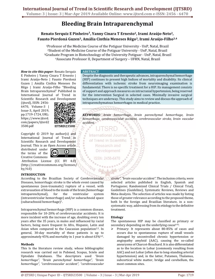
International Journal of Trend in Scientific Research and Development (IJTSRD) Volume: 3 | Issue: 3 | Mar-Apr 2019 Available Online: www.ijtsrd.com e-ISSN: 2456 - 6470 Bleeding Brain Intraparenchymal Renato Serquiz E Pinheiro¹, Yanny Cinara T Ernesto2, Irami Araújo-Neto2, Fausto Pierdoná Guzen3, Amália Cinthia Meneses Rêgo3, Irami Araújo-Filho3,4 1Professor of the Medicine Course of the Potiguar University - UnP, Natal, Brazil 2Student of the Medicine Course of the Potiguar University - UnP, Natal, Brazil 3Graduate Program in Biotechnology of the University Potiguar - UnP, Natal, Brazil 4Associate Professor II, Department of Surgery – UFRN, Natal, Brazil How to cite this paper: Renato Serquiz E Pinheiro | Yanny Cinara T Ernesto | Irami Araújo-Neto | Fausto Pierdoná Guzen | Amália Cinthia Meneses Do Rêgo | Irami Araújo-Filho "Bleeding Brain Intraparenchymal" Published in International Journal of Trend in Scientific Research and Development (ijtsrd), ISSN: 2456- 6470, Volume-3 | Issue-3, April 2019, pp.1719-1724, URL: https://www.ijtsrd. com/papers/ijtsrd2 3500.pdf Copyright © 2019 by author(s) and International Journal of Trend in Scientific Research and Development Journal. This is an Open Access article distributed under the terms of the Creative Commons Attribution License (CC BY 4.0) (http://creativecommons.org/licenses/ by/4.0) INTRODUCTION According to the Brazilian Society of Cerebrovascular Diseases, hemorrhagic stroke is the whole event caused by spontaneous (non-traumatic) rupture of a vessel, with extravasation of blood to the inside of the brain (hemorrhage intraparenchymal), for the ventricular system (intraventricular hemorrhage) and/or subarachnoid space (subarachnoid hemorrhage)1. Intraparenchymal hemorrhage (HIP) is a common disease, responsible for 10-20% of cerebrovascular accidents. It is more incident with the increase of age, doubling every ten years after the 35 years, in males and influenced by racial factors, being more frequent In Afro, Hispanic, Latin and Asian when compared to the Caucasian population2,3. In general, 30-day mortality of these patients is up to approximately 45% and mortality in 1 year is about 63%4,5. Methods This Is the literature review study, whose bibliographic research was carried out in Pubmed, Scopus, Scielo and Uptodate Databases. The descriptors used “brain hemorrhage”, “brain parenchymal hemorrhage”, “brain hemorrhage”, “cerebrovascular accident”, “cerebrovascular ABSTRACT Despite the diagnostic and therapeutic advances, intraparenchymal hemorrhage (HIP) continues to present high Indices of mortality and disability. Its clinical differentiation with ischemic stroke from neuroimaging examination is fundamental. There is no specific treatment for a HIP. Its management consists of support and approach measures on intracranial hypertension, being reserved for the intervention Surgical in selected cases. Minimally invasive surgical techniques are underway. This study aims to review and discuss the approach of intraparenchymatous hemorrhages in medical practice. KEYWORDS: brain hemorrhage, brain parenchymal hemorrhage, brain hemorrhage, cerebrovascular accident, cerebrovascular stroke, brain vascular accident. stroke”, “brain vascular accident”. The inclusion criteria, were selected articles published in English, Spanish and Portuguese; Randomized Clinical Trials / Clinical Trial), Guidelines (Guideline), Systematic Reviews, Reviews and Meta-Analysis. The selection of articles was made by listing those of greater relevance according to the proposed theme, both In the foreign and Brazilian literature, in a non- systematic way, addressing from its etiology to the definitive treatment. Etiology The spontaneous HIP may be classified as primary or secondary depending on the underlying cause6-8: ?Primary: It represents about 80-85% of cases and occurs due to spontaneous rupture of small vessels damaged by uncontrolled chronic hypertension or angiopathy amyloid (AAC), causing the so-called aneurysms of Charcot-Bouchard. It is also differentiated as to the location in Lobar (commonly resulting from AAC) and not Lobar (often due to long-standing arterial hypertension) and, in the latter, Putamen, Thalamus, subcortical white matter, bridge and cerebellum, the most common sites. IJTSRD23500 @ IJTSRD | Unique Paper ID – IJTSRD23500 | Volume – 3 | Issue – 3 | Mar-Apr 2019 Page: 1719
International Journal of Trend in Scientific Research and Development (IJTSRD) @ www.ijtsrd.com eISSN: 2456-6470 ?Secondary: Corresponds to 15-25% of cases and is associated with a series of congenital and acquired conditions, such as vascular pathologies (arteriovenous malformations, Moyamoya, Cavernomas, Venous Angiomas and Telangiectasis), tumors, hemorrhagic transformation of cerebral ischemia, leukemia, coagulation disorders, use of anticoagulants and thrombolytic agents, cerebral vasculitis, drug abuse, cerebral venous thrombosis, among other causes. Risk Factors Non-modifiable ?Advanced age; ?Black and Oriental breed; ?Male. Modifiable ?Hypertension: is the main risk factor, being present in 70 to 80% of patients with HIP; ?Angiopathy Amyloid: is characterized by deposits of the beta peptide Amyloid within small and medium vessels of the cortex and leptomeningeal. It may occur as a sporadic disorder, sometimes in association with Alzheimer's disease, or as a specific familial syndrome. It is a significant cause of HIP lobar primary in the elderly, especially in the parietal and occipital lobes; ?Smoking: Smokers have a higher risk of stroke, including HIP than in non-smokers; ?Alcohol: Consumption of moderate or large quantities of alcohol in the 24h preceding the ictus seems to precipitate HIP; ?Coagulopathies: Both the coagulopathies primary and secondary, such as the use of oral anticoagulants, antiplatelet drugs, thrombolytics for the treatment of acute ischemic stroke or acute myocardial infarction (AMI) are associated with increased risk of the HIP; ?Sympathomimetics Medicines or drugs with sympathomimetic activity, (Phenylpropanolamine, cocaine, amphetamines, or ephedrine) also increase the risk of the HIP; ?Cholesterol and triglyceride Status: There may be an inverse relationship between the levels of total cholesterol, LDL and triglycerides and the risk of a HIP. ?Other: Less established factors, such as obesity and genetic factors, are also pointed out as relevant to the risk of the HIP. Clinical picture HIP is usually characterized by the sudden focal neurological deficit, dependent on the bleeding topography, sudden onset and rapidly progressive. Although frequent, the headache is not present in all cases, only when the HIP is accompanies of meningeal irritation due to intraventricular hemorrhage or subarachnoid associated, or increased intracranial pressure9. For this last reason or by distortion of brain structures, the vomiting is associated. Convulsive seizures may occur, especially in bleeding Lobar10. Bleeding in the Putamen occurs in approximately 35% of the cases, subcortical in 30%, cerebellum in 16%, thalamus in 15% and bridge in 5-12%11-13. They manifest themselves with: ?Putamen: Hemiplegia, loss hemisensory, hemianopsia homonymous, gaze paralysis, torpor, and coma. ?Cerebellar: Imbalance, vomiting, headache (usually occipital), stiffness nuchal, paralysis of the gaze, facial paresis or even torpor due to compression of the brainstem. It is a crucial diagnosis since these patients frequently deteriorate and require surgery. ?Thalamic: hemiparesis, occasionally hemianopsia transient homonymous, and there may be pupils miotic non-reactive. Aphasia can occur if bleeding affects the dominant hemisphere or neglect in the non-dominant hemisphere. ?Pontina: Usually leads to deep coma in the first few minutes after hemorrhage, probably due to rupture of the reticular activation system. The motor examination is marked by total paralysis. The pupils are reactive, but the ocular movements are absent, and there may be ocular oscillation, facial paralysis, deafness, and dysarthria. When the patient is awake. It is also essential that all patients with suspected HIP are evaluated by the Glasgow Coma Scale and by NIHSS (National Institutes Of Heath Stroke Scale) in the evaluation neurological. By hard clinical differentiation with ischemic stroke, a neuroimaging test for diagnostic confirmation of HIP is essential14-16. Complementary exams In addition to a rapid clinical history and neurological examination, computed tomography without hire (TCSC) of the skull is highly sensitive and specific for hemorrhagic stroke and is the key to early diagnosis17. In the HIP, it will reveal not only the location and size of the hemorrhage but also the intraventricular extension, the mass effect, the hydrocephalus and the first signs of herniation. The volume of the hematoma can be estimated using the ABC/2 method, where6-9,18: ?A - Is the maximum diameter of the hematoma (in cm); ?B - Is the largest perpendicular diameter A (in cm); ?C - Is the sum of the points obtained with the 10mm cuts, in which the main axis of the hematoma presents: ?75% to 100% of a = 1 point for each cut; ?25% to 50% of a = 0.5 point each cut; ?≤ 25% of a = Do not score; Hematoma Volume (mL) = A x B x C / 2 This calculation of the hematoma volume, although variable among the examiners, proved to be sufficient for clinical decision making since hematomas with more than 30mL have a worse prognosis. MRI can be as sensitive as CCTSC to determine the presence of hemorrhage, but rarely offers additional information in the acute situation, and may be performed when there is suspicion of non-hypertensive etiology (Cavernomas, Angiopathy Amyloid, Neoplasms, among others). As a general rule, the hyperacute hematoma is isointense in T1, hyperintense in T2 and hypointense in T2. After the seventh day, it becomes hyperintense on T1 and T2 due to hemoglobin degradation in Methemoglobin, and in the chronic phase, the low signal of hemosiderin is better visualized in T2 or T219. Angiography is indicated for investigation of secondary causes such as AVM, aneurysms, fistulas, venous sinus thrombosis and vasculitides in patients with atypical HIP localization or aged below 45 years (regardless of the presence of SAH). The method of choice is conventional catheter angiography, but CT or MRI angiography with use of loss hemisensory and @ IJTSRD | Unique Paper ID - IJTSRD23500 | Volume – 3 | Issue – 3 | Mar-Apr 2019 Page: 1720
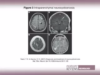
International Journal of Trend in Scientific Research and Development (IJTSRD) @ www.ijtsrd.com eISSN: 2456-6470 Gadolinium are less invasive alternatives with good sensitivity (Figure 1)12. respiratory and hemodynamic parameters, temperature, capillary glycemia and detection of focal and external neurological signs of trauma and its complications21. Airway stabilization, respiration, and circulation (ABC) are essential to prevent secondary injuries caused by hypoxemia, uncontrolled hypertension, and hematoma expansion. Intubation for airway protection is indicated in patients with Glasgow Scale (GCS) ≤8 or significant respiratory distress. Ideally, after performing the neuroimaging exam, patients with HIP should be quickly referred to beds monitored in a stroke unit or intensive care beds10-13. Temperature Fever is an independent risk factor for poor prognosis. In about 20% of patients, there is no infection and fever is attributed to inflammatory responses to extravasated blood, predominating in those with a poor clinical condition at admission and in those with an intraventricular extension of hemorrhage7-9. It is suggested that hyperthermia (axillary temperature ≥37.5ºC) is treated in the acute phase of HIP usually with acetaminophen or dipyrone. In refractory cases, it may be associated with anti-inflammatory non-steroidal, external physical methods and internal active cooling (intravascular cooling catheters), although there are no studies proving efficacy and safety of cooling in patients with hemorrhagic stroke1-3. Glucose Care should be taken to avoid hyperglycemia and hypoglycemia. Insulin therapy is recommended in order to reach blood glucose levels between 100-140mg/dL For patients with hemorrhagic stroke16,17. Prophylaxis for venous thromboembolism (VTE) There is a significant risk of VTE in patients with hemorrhagic stroke. Therefore, in patients with HIP restricted to bed, it is recommended to start the mechanical prophylaxis of VTE, preferably with intermittent pneumatic compression devices, or if not available, compression stockings graduated at the time of admission. Associated with mechanical prophylaxis, the prophylactic doses of non- fractioned subcutaneous heparin or LMWH may be initiated after the first 48h in patients with documented stable hematoma22,23. Blood pressure (BP) In the HIP, the elevation of systolic blood pressure (SBP) is associated with the expansion of the hematoma, neurological deterioration, and lower prognosis. Thus, treatment is suggested if SBP>180mmHg and mean BP (map) > 130mmHg, aiming to reach a BP close to 160x90mmHg (or Pam <110 mmHg)18-20. In patients with intracranial hypertension, SBP should be maintained above 90mmHg and ideally, intracranial pressure (PIC) monitoring should be used to maintain cerebral perfusion pressure (CPP) above 60–80mmHg (PPC = PAM – PIC)6. The medications antihypertensive generally used in the acute phase of the HIP are Metoprolol, Diltiazem or Esmolol since the Labetalol And Nicardipine are not available for use in Brazil (Table 3). For more severe or refractory cases, it is recommended to Nitroprusside of sodium IV, paying attention to a potential increase in ICP. Oral anti- hypertensive medicinal products should be instituted as soon as possible (Tables 2 - 3)1. Figure1. The typical locations of hypertensive HIP are: Putamen (A), thalamus (B), white matter Subcortical (C), Bridge (D) and cerebellum (e). Thalamic hemorrhages and Subcortical Frequently Extend to the ventricles (B and C). ADrug search or vascular anomaly often causes bleeding Lobar (F). Source: Dastur et al, 2017. Prognosis Despite the diagnostic and therapeutic advances, HIP continues to present high Indices of mortality and disability. The main factors related to the poor prognosis in the short term (in 30 days) of these patients are initial volume of hemorrhage more significant than 30cm³, lowering of the level of consciousness at admission, intraventricular bleeding, advanced age, and localization primarily Infratentorial20. For stratification of the risk of mortality in 30 days, the HIP scale is the most used. Scores between 0 and 2 are associated with low mortality and scores ≥ 3 are associated with a high mortality rate (Table 1)7,11. Table1. HIP Scale Components Glasgow Scale 3-4 5-12 13-15 Hematoma Volume (cm³) ≥ 30 < 30 Ventricular flooding Yes No Origin Infratentorial Yes No Age (years) ≥ 80 < 80 Total Source: Hemphill et al. (2001) Treatment There is no specific treatment for the HIP. The initial approach should be focused on airway evaluation, Points 2 1 0 1 0 1 0 1 0 1 0 0-6 @ IJTSRD | Unique Paper ID - IJTSRD23500 | Volume – 3 | Issue – 3 | Mar-Apr 2019 Page: 1721
International Journal of Trend in Scientific Research and Development (IJTSRD) @ www.ijtsrd.com eISSN: 2456-6470 Table2. Blood pressure control in HIP Intracranial hypertension Increased intracranial pressure (ICP/PIC) due to HIP may result from the hematoma itself and the surrounding edema, which may contribute to brain injury and neurological deterioration. A graded approach is recommended starting with simple measures that include7-9: ?Elevation of the headboard in 30⁰ and positioning of the head, avoiding excessive flexion or rotation of the neck; ?Analgesia (Morphine or Fentanyl) and Sedation (Propofol, Etomidate Or Midazolam) particularly in unstable and intubated patients; ?Keep the Euvolemia: If necessary, isotonic fluids (e.g., saline solution 0.9%) should be administered. ?BP control; ?Treatment of fever and seizures; Invasive monitoring and specific treatment of PIC should be considered for patients with GCS<8, with clinical evidence of herniation transtentorial or with significant intraventricular hemorrhage or hydrocephalus12. The goal should be to keep the PIC< 20mmHg. More aggressive therapies to obtain this control include osmotic diuretics (e.g., mannitol and hypertonic saline solution), neuromuscular blockade, hyperventilation, hypothermia, and Ventriculostomy 10,13-15. ?Mannitol 20%: bolus Initial dose of 1g/Kg, followed by infusions from 0.25 to 0.5 g/Kg/dose (IV) administered for 20min, from 6/6 hours (usual maximum dosage: 2 g/Kg); ?Hypertonic Saline Solution: NaCl 3%, in continuous infusion, with control of serum sodium every 6h, respecting the maximum increase of sodium around 15 mEq/L/day, in refractory cases; ?Analgesia and/or neuromuscular barbiturates (phenobarbital 5-20 mg/Hg in bolus followed by 1-4mg/Kg/h) associated or not with non- depolarizing neuromuscular blockers; ?Hyperventilation: The objective is to reduce the PaC02 to levels between 25-30 mmHg, determining almost immediate cerebral vasoconstriction. It causes a reduction of 25%-30% in intracranial pressure, with maximum effect in approximately thirty minutes. ?Induced hypothermia: through the cooling of all the body, its objective is to maintain a temperature of 32- 34oC, but its therapeutic use in the elevated PIC remains controversial and should be limited to refractory patients. ?Ventriculostomy: indicated hydrocephalus (a common complication of thalamic hemorrhage with compression of the III ventricle and cerebellar hemorrhage with compression of the IV ventricle) or intraventricular hemorrhage with hydrocephalus. Reversal of Coagulopathies Approximately 12-20% of patients with HIP make use of oral anticoagulants. In this way, the rapid correction of the coagulopathy should be considered in any potentially recoverable patient, and any antithrombotic agent should be discontinued immediately. The method of reversal will depend on the agent used and should not be postponed until the results of the laboratory coagulation tests have arrived24. If you use warfarin: immediately administer fresh frozen plasma (FFP) or complex concentrate Prothrombin (CCP) and vitamin K (IV) 10mg until normalization of prothrombin time measured by means of INR<1.4. CCP is an inactivated Blood pressure Conduct Initiate aggressive BP reduction by continuous infusion of intravenous antihypertensive medication with BP monitoring every 5 minutes. Consider ICP monitoring. Initiate BP reduction by continuous or intermittent infusion of intravenous antihypertensive medication with BP monitoring every 5 minutes. Keep PPC>60- 80mmHg. Initiate moderate BP reduction by continuous or intermittent infusion of antihypertensive With BP monitoring every 15 minutes (target BP 160x90mmhg or PAM target 110mmhg). Expansion with Crystalloids intravenously and infusion of vasoactive amines: Dopamine 2 – 20mg/Kg/min. / noradrenaline 0.05 – 0.2 mg/Kg/min. SBP >200 mmHg or PAM>150 mmHg (two readings with a 5- minute interval) PAS>180 mmHg or PAM>130mmHg Suspected increase in ICP PAS>180 mmHg or PAM>130 mmHg No suspected increase in ICP PAS<90mmHg BP: blood pressure; PAM: mean arterial pressure; ICP: Intracranial pressure; PPC: Cerebral perfusion pressure source: Pontes-Neto et al. (2009). Table3. Antihypertensive medications used in HIP Intravenous Dose 5mg-1ML/min every 10 min, up to a maximum of 20 min. blockade: Drug Contraindications Severe HF, COPD, asthma, hypotension, bradycardia Sudden BP drop in renin-increasing states. ARF if renal artery stenosis. Metoprolol 0,625-1.25 mg in 5min every 6h. Enalapril 0.25-0.35mg/kg at 10min Infusion 5-15 mg/h. in obstructive Sinus node disease or atrioventricular node. Severe HF Diltiazen Potential increase in ICP, variable response, cyanide poisoning and Thiocyanate. Nitroprusside Sodium 0.25-10 mg/Kg/min. 250- 500mg/Kg/min in bolus every 10min or infusion 25- 300mg/Kg/min Severe HF, COPD, asthma, hypotension, bradycardia. Esmolol HF: Heart failure; ARF: acute renal failure; COPD: Chronic obstructive pulmonary disease; BP: blood pressure; ICP: Intracranial pressure. Source: Modified de Pontes-Neto et al. (2009) @ IJTSRD | Unique Paper ID - IJTSRD23500 | Volume – 3 | Issue – 3 | Mar-Apr 2019 Page: 1722
International Journal of Trend in Scientific Research and Development (IJTSRD) @ www.ijtsrd.com eISSN: 2456-6470 concentrate of factors II, IX and X, with variable amounts of factor VII, its concentration of coagulation factors dependent on vitamin K is about 25 times greater than that of fresh frozen plasma (FFP) and can reverse the coagulopathy rapidly, with lower volume and lower risk of infection, pulmonary edema, transfusion-related acute lung injury (Trali) and cardiac overload associated with transfusion (COAT)9-11. If the use of unfractionated heparin: administer sulfate Protamine (IV) 1mg for each 100units of heparin administered in the last 2-3 hours (maximum 50mg). Repeat half of the initial dose if ttpa remains elevated3. If symptomatic HIP associated with the use of thrombolytics: administer fresh frozen plasma, cryoprecipitate, and platelets18. Seizures HIP patient has up to 16% risk of clinical seizures within a week, Particularly In those where there is hemorrhage Lobar With intraventricular flooding. Therefore, prophylactic treatment with anticonvulsants is recommended in patients with torporous and comatose with activity Epileptiform Electroencephalogram (EEG) in patients with bleeding Lobar and in those where there are signs of intracranial hypertension25. The drugs of choice are Phenytoin and Phenobarbital, which should be maintained at therapeutic serum levels for one month and subsequently withdrawn gradually14. Surgery Surgical interventions to approach the mass effect by the hematoma include Craniotomy Decompressive of the posterior fossa or drainage of the hematoma and are indications6,18: ?Cerebellar Hematoma >3cm in diameter with deterioration brain stem hydrocephalus; ?Bleeding Lobar voluminous and in up to 1cm from the surface of the cerebral cortex, especially in young patients, with scores on the Glasgow coma scale between nine and twelve. In cases of hydrocephalus, an external ventricular shunt may be necessary during the critical period of ICP elevation26. The Craniotomy Is the most studied surgical technique in patients with HIP supratentorial. However, other methods include endoscopic aspiration of the hemorrhage, fibrinolytic therapy to undo the clot followed by aspiration and aspiration stereotactic guided by CT. Studies of these less invasive techniques are underway27,28. Conclusion The acute treatment of intraparenchymal cerebral hemorrhage is still a subject that remains in an intense investigation. The lack of guidelines and more current guidelines in Brazil evidences the need for scientific reviews with explicit language, which seek to standardize care and practical approach to patients who are affected by this disease. Although there is no consensus yet, the multiple ongoing studies show evidence of efficacy in reducing blood pressure and redefining new targets. Recent neuroimaging studies already reveal patterns that identify patients who benefit most from minimally neuroprotection studies in surrounding areas of the lesion confirm the benefit of correct measures of intracranial hypertension, glycemic control, and body temperature. The treatment of intracranial hemorrhage continues to be directed towards the reduction of disability and morbidity and mortality. However, prevention measures are widely studied and disseminated in clinical practice. In this area of prevention, adequate blood pressure control is of paramount importance. References [1]Pontes-Neto, Octávio M. et al. Diretrizes para o manejo de pacientes com hemorragia intraparenquimatosa cerebral espontânea. Arq. de Neuro-psiquiatria, [s.l.], v. 67, n. 3, p.940-950, 2009. doi:10.1590/s0004- 282x2009000500034. invasive surgeries. And [2] Fewel ME, Thompson BG Jr, Hoff JT. Spontaneous intracerebral hemorrhage: a review. Neurosurg Focus. 2003 Oct 15;15(4):E1. [3] Ikram MA, Wieberdink RG, Koudstaal PJ. International epidemiology of intracerebral hemorrhage. Curr Atheroscler Rep. 2012 Aug; 14(4):300-6. doi: 10.1007/s11883-012-0252-1. [4] Tellería-Díaz A. [Management and predictors in patients with spontaneous intracerebral hemorrhage]. Rev Neurol. 2006 Mar 16-31; 42(6):341-9. [5]Rordorf, intracerebral hemorrhage: Pathogenesis, clinical features, and diagnosis. Uptodate. Feb, 2017. In: https:// uptodate.com/contents/spontaneous- intracerebral-hemorrhage-pathogenesis features-and-diagnosis. Guy; Mcdonald, Colin. Spontaneous clinical- compression and/or [6] Labovitz DL, Sacco RL. Intracerebral hemorrhage: update. Curr Opin Neurol. 2001 Feb; 14(1):103-8. [7]Hemphill, J. C. et al. The ICH Score: A Simple, Reliable Grading Scale for Intracerebral Hemorrhage Editorial Comment. Stroke, [s.l.], v. 32, n. 4, p.891-897, 2001. Ovid Technologies (Wolters doi:10.1161/01.str.32.4.891. Kluwer Health). [8] Morioka M, Orito K. Management of Spontaneous Intracerebral Hematoma. Neurol Med Chir (Tokyo). 2017 Nov 15; 10.2176/nmc.ra.2016-0327. 57(11):563-574. doi: [9]Wellman GC, Koide M. Impact of subarachnoid hemorrhage on parenchymal arteriolar function. Acta Neurochir Suppl. 2013; 115:173-7. doi: 10.1007/978- 3-7091-1192-5_33. [10] Kang J, Huang Q, Liu Y. Advance in research on the genetic etiology of spontaneous intracerebral hemorrhage. Zhonghua Yi Xue Yi Chuan Xue Za Zhi. 2016 Oct; 33(5):702-7. doi: 10.3760/cma.j.issn.1003- 9406.2016.05.028. [11]Hemphill, J. Claude et al. Guidelines for the Management of Spontaneous Hemorrhage. Stroke, [s.l.], v. 46, n. 7, p.2032-2060, 2015. Ovid Technologies (Wolters Kluwer Health). doi:10.1161/str.0000000000000069. Intracerebral @ IJTSRD | Unique Paper ID - IJTSRD23500 | Volume – 3 | Issue – 3 | Mar-Apr 2019 Page: 1723
International Journal of Trend in Scientific Research and Development (IJTSRD) @ www.ijtsrd.com eISSN: 2456-6470 [12]Dastur, Cyrus K; YU, Wengui. Current management of spontaneous intracerebral haemorrhage. Bmj, [s.l.], p.000047-000058, 2017. BMJ. doi: 10.1136/svn-2016- 000047. [21] Touma L, Filion KB, Sterling LH, Atallah R, Windle SB, Eisenberg MJ. Stent Retrievers for the Treatment of Acute Ischemic Stroke: A Systematic Review and Meta- analysis of Randomized Clinical Trials. JAMA Neurol. 2016 Mar; 10.1001/jamaneurol.2015.4441. 73(3):275-81. doi: [13] Domingues R, Rossi C, Cordonnier C. Diagnostic evaluation for nontraumatic intracerebral hemorrhage. Neurol Clin. 2015 10.1016/j.ncl.2014.12.001. [22] Sun H, Zhang H, Ma J, Liu Y, You C. Evaluating the diagnostic accuracy of CT perfusion in patients with cerebral vasospasm after aneurysm rupture: a meta- analysis. Turk Neurosurg. 2014; 24(5):757-62. doi: 10.5137/1019-5149.JTN.9914-13.1. May; 33(2):315-28. doi: [14]Coletto, Francisco Antônio et al. Manual de rotinas para atenção ao AVC. Brasília: Editora do Ministério da Saúde, 2013. 54 bvsms.saude.gov.br/bvs/publicacoes/manual_rotinas_ para_atencao_avc.pdf p. Disponível em: [23] Whiteley WN, Slot KB, Fernandes P, Sandercock P, Wardlaw J. Risk factors for intracranial hemorrhage in acute ischemic stroke patients treated with recombinant tissue plasminogen activator: a systematic review and meta-analysis of 55 studies. Stroke. 2012 Nov; 43(11):2904-9. 10.1161/STROKEAHA.112.665331. [15]Greenberg, Steven M; Cerebral amyloid angiopathy. Uptodate. Feb, uptodate.com/contents/cerebral-amyloid-angiopathy 2017. In: https:// doi: [16]Rordorf, intracerebral hemorrhage: Treatment and prognosis. Uptodate. Feb, https://uptodate.com/contents/spontaneous- intracerebral-hemorrhage-treatment-and-prognosis. Guy; Mcdonald, Colin. Spontaneous [24] Hacke W, Donnan G, Fieschi C, Kaste M, von Kummer R, Broderick JP, Brott T, Frankel M, Grotta JC, Haley EC Jr, Kwiatkowski T, Levine SR, Lewandowski C, Lu M, Lyden P, Marler JR, Patel S, Tilley BC, Albers G, Bluhmki E, Wilhelm M, Hamilton S; ATLANTIS Trials Investigators; ECASS Trials Investigators; NINDS rt-PA Study Group Investigators. Association of outcome with early stroke treatment: pooled analysis of ATLANTIS, ECASS, and NINDS rt-PA stroke trials. Lancet. 2004 Mar 6; 363(9411):768-74. 2017. In: [17]Smith, Edward R; Amin-Hanjani, Sepideh. Evaluation and management of elevated intracranial pressure in adults. Uptodate. Feb, uptodate.com/contents/evaluation-and-management- of-elevated-intracranial-pressure-in-adults 2017. In: https:// [18]Fiorella, Dave et al. Intracerebral Hemorrhage: A Common and Devastating Disease in Need of Better Treatment. World Neurosurgery, [s.l.], v. 84, n. 4, p.1136-1141, out. doi:10.1016/j.wneu.2015.05.063. [25] Coutinho JM, Liebeskind DS, Slater LA, Nogueira RG, Clark W, Dávalos A, Bonafé A, Jahan R, Fischer U, Gralla J, Saver JL, Pereira VM. Combined Intravenous Thrombolysis and Thrombectomy vs Thrombectomy Alone for Acute Ischemic Stroke: A Pooled Analysis of the SWIFT and STAR Studies. JAMA Neurol. 2017 Mar 1; 74(3):268-274. doi: 10.1001/jamaneurol.2016.5374. 2015. Elsevier BV. [19]Miranda, Renata et al. Diretriz de acidente vascular cerebral do Hospital Israelita Albert Einstein. São Paulo: HIAE, 2013. pubdiretrizes.einstein.br/download.aspx?ID=%7BD86 6950D-5445-4BC2-833D 6994487DB40D%7D. 130 p. Disponível em: [26] Bereczki D, Liu M, Prado GF, Fekete I. Cochrane report: A systematic review of mannitol therapy for acute ischemic stroke and hemorrhage. Stroke. 2000 Nov; 31(11):2719-22. cerebral parenchymal [20] Goyal M, Menon BK, van Zwam WH, Dippel DW, Mitchell PJ, Demchuk AM, Dávalos A, Majoie CB, van der Lugt A, de Miquel MA, Donnan GA, Roos YB, Bonafe A, Jahan R, Diener HC, van den Berg LA, Levy EI, Berkhemer OA, Pereira VM, Rempel J, Millán M, Davis SM, Roy D, Thornton J, Román LS, Ribó M, Beumer D, Stouch B, Brown S, Campbell BC, van Oostenbrugge RJ, Saver JL, Hill MD, Jovin TG; HERMES collaborators. Endovascular thrombectomy ischaemic stroke: a meta-analysis of individual patient data from five randomised trials. Lancet. 2016 Apr 23; 387(10029):1723-31. 6736(16)00163-X. [27] Sadaka F, Kasal J, Lakshmanan R, Palagiri A. Placement of intracranial pressure monitors by neurointensivists: case series and a systematic review. Brain Inj. 2013; 27(5):600-4. doi: 10.3109/02699052.2013.772238. [28] Kang J, Huang Q, Liu Y. Advance in research on the genetic etiology of spontaneous intracerebral hemorrhage. Zhonghua Yi Xue Yi Chuan Xue Za Zhi. 2016 Oct; 33(5):702-7. doi: 10.3760/cma.j.issn.1003- 9406.2016.05.028. after large-vessel doi: 10.1016/S0140- @ IJTSRD | Unique Paper ID - IJTSRD23500 | Volume – 3 | Issue – 3 | Mar-Apr 2019 Page: 1724