Download
1 / 4
40 likes | 48 Views
The ubiquitin proteasome system UPS is essential for many cellular processes, including the cell cycle, the regulation of gene expression and cell survival. Dysfunctional UPS can be associated with the underlying pathophysiology of specific diseases. The 20S proteasome core is composed of 28 subunits, which are arranged in four stacked rings, resulting in a barrel shaped structure. The two end rings are each formed by seven subunits, and the two central rings are each formed by seven subunits. The The over expression of LMP2 1i in trophoblast cells of hydatidiform moles may contribute to its highly invasive phenotype. LMP2 1i deficient mice reportedly exhibit uterine neoplasms, with a disease prevalence of 36 by 12 months of age. Embryo implantation involves the invasion of placental extravillous trophoblast cells EVTs into the uterus. Normal human placentas or placentas from hydatidiform mole patients were collected and the expression of LMP2 1i in different cell types including trophoblastic column TC , cytotrophoblast cells CTB and syncytiotrophoblasts STBs was examined under different pathological states by pathological analysis. The expression of LMP2 1i in TC of partial hydatidiform mole and complete hydatidiform mole placentas, was higher than that in TC of normal human placentas. Further the experiments with human and mouse uterine tissues clarified the physiological significance of LMP2 1i in malignant myometrium transformation. In this mini review, we covered recent insights into the molecular pathways involved in LMP2 1i mediated physiological functions, with a particular focus on embryo implantation and uterine mesenchymal tumorigenesis. Takuma Hayashi | Kenji Sano | Tomoyuki Ichimura | Gal Gur | Hiroyuki Aburatani | Yae Kanai | Dorit Zharhary | Susumu Tonegawa | Nobuo Yaegashi | Ikuo Konishi "Physiological Functions of LMP2/B1i in the Female Reproductive System" Published in International Journal of Trend in Scientific Research and Development (ijtsrd), ISSN: 2456-6470, Volume-3 | Issue-4 , June 2019, URL: https://www.ijtsrd.com/papers/ijtsrd23892.pdf Paper URL: https://www.ijtsrd.com/medicine/physiology/23892/physiological-functions-of-lmp2b1i-in-the-female-reproductive-system/takuma-hayashi<br>

E N D
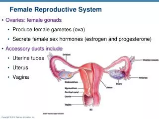
International Journal of Trend in Scientific Research and Development (IJTSRD) Volume: 3 | Issue: 4 | May-Jun 2019 Available Online: www.ijtsrd.com e-ISSN: 2456 - 6470 Physiological Functions of LMP2/β β β β1i in the Female Reproductive System Takuma Hayashi1,9,10, Kenji Sano2, Tomoyuki Ichimura3, Gal Gur4,10, Hiroyuki Aburatani5, Yae Kanai6, Dorit Zharhary4,10, Susumu Tonegawa7, Nobuo Yaegashi8, Ikuo Konishi1 1National Hospital Organization Kyoto Medical Center, Japan 2Department of Laboratory Medicine, Shinshu University Hospital, Nagano, Japan 3Department of Obstetrics and Gynecology, Osaka City University Graduate School of Medicine, Osaka, Japan 4Sigma-Aldrich Israel Ltd., Rehovot, Israel 5The Cancer System Laboratory, Research Center for Advanced Science and Technology, The University of Tokyo, Japan 6Pathology Division, Keio University School of Medicine, Tokyo 7Picower Institution and Department of Biology, Massachusetts Institute of Technology, MA, USA 8Department of Obstetrics and Gynecology, Tohoku University Graduate School of Medicine, Miyagi, Japan 9Promoting Business using Advanced Technology, Japan Science and Technology Agency (JST), Tokyo, Japan 10SIGMA-Aldrich Collaboration Laboratory How to cite this paper: Takuma Hayashi | Kenji Sano | Tomoyuki Ichimura | Gal Gur | Hiroyuki Aburatani | Yae Kanai | Dorit Zharhary | Susumu Tonegawa | Nobuo Yaegashi | Ikuo Konishi "Physiological Functions of LMP2/B1i in the Female Reproductive System" Published in International Journal of Trend in Scientific Research and Development (ijtsrd), ISSN: 2456- 6470, Volume-3 | Issue-4, June 2019, pp.597-600, URL: https://www.ijtsrd.c om/papers/ijtsrd23 892.pdf Copyright © 2019 by author(s) and International Journal of Trend in Scientific Research and Development Journal. This is an Open Access article distributed under the terms of the Creative Commons Attribution License (CC BY 4.0) (http://creativecommons.org/licenses/ by/4.0) INTRODUCTION The eukaryotic UPS is responsible for most aspects of regulatory and quality-control protein degradation in cells. Its substrates, which are usually modified by polymers of ubiquitin, are ultimately degraded by the immuno- proteasome [1,2]. The ubiquitin-proteasome system (UPS) controls almost all basic cellular processes, such as progression through the cell cycle, signal transduction, cell death, immune responses, metabolism, protein quality control and development by degrading short-lived regulatory or structurally aberrant proteins [1,3,4]. Cytoplasmic proteins are mostly degraded by a protease complex, which has many substrates consisting of twenty- eight 20 to 30-kDa subunits, referred to as the immuno- ABSTRACT The ubiquitin proteasome system (UPS) is essential for many cellular processes, including the cell cycle, the regulation of gene expression and cell survival. Dysfunctional UPS can be associated with the underlying pathophysiology of specific diseases. The 20S proteasome core is composed of 28 subunits, which are arranged in four stacked rings, resulting in a barrel-shaped structure. The two end rings are each formed by seven α subunits, and the two central rings are each formed by seven β subunits. The The over expression of LMP2/β1i in trophoblast cells of hydatidiform moles may contribute to its highly invasive phenotype. LMP2/β1i-deficient mice reportedly exhibit uterine neoplasms, with a disease prevalence of 36% by 12 months of age. Embryo implantation involves the invasion of placental extravillous trophoblast cells (EVTs) into the uterus. Normal human placentas or placentas from hydatidiform mole patients were collected and the expression of LMP2/β1i in different cell types including trophoblastic column (TC), cytotrophoblast cells (CTB) and syncytiotrophoblasts (STBs) was examined under different pathological states by pathological analysis. The expression of LMP2/β1i in TC of partial hydatidiform mole and complete hydatidiform mole placentas, was higher than that in TC of normal human placentas. Further the experiments with human and mouse uterine tissues clarified the physiological significance of LMP2/β1i in malignant myometrium transformation. In this mini review, we covered recent insights into the molecular pathways involved in LMP2/β1i-mediated physiological functions, with a particular focus on embryo implantation and uterine mesenchymal tumorigenesis. Keywords: LMP2/β1i, implantation, trophoblast, leiomyosarcoma, leiomyoma IJTSRD23892 proteasome, and it plays key roll functions in the nucleus and cytoplasm of eukaryotic cells, while LMP2/β1i and LMP7/β5i individually appear to be more intense in the endoplasmic reticulum [5]. The proteasome structure is a cylindrical complex containing a core of four stacked rings around a central pore, with each ring being composed of seven individual proteins. The inner two rings are made of seven β subunits that contain three to seven protease active sites [6-10]. Two of the βsubunits with an NH2-terminal threonine residue, low molecular mass polypeptide (LMP) 2/β1i and LMP7/σ5ι, which are induced by interferon (IFN)- γ, are encoded within the class II region of the MHC, directly adjacent to the transporter associated with antigen @ IJTSRD | Unique Paper ID - IJTSRD23892 | Volume – 3 | Issue – 4 | May-Jun 2019 Page: 597
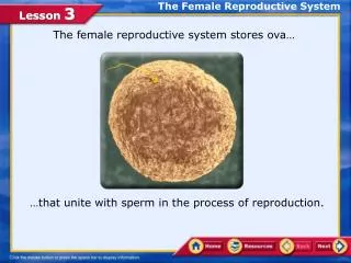
International Journal of Trend in Scientific Research and Development (IJTSRD) @ www.ijtsrd.com eISSN: 2456-6470 presentation (TAP) 1 and TAP2 genes [11]. Several experiments have shown that IFN-γ-induced-incorporation of LMP2/β1i and LMP7/β5i into the 20S proteasome is responsible for antigen presentation [12,13]. Furthermore, the proteasome reconstructed by LMP2/β1i and LMP7/β5i, referred to as immuno-proteasome, produced increased chymotryptic and tryptic protease activities and modulated cleavage-site preferences of the proteasome [14-17]. Thus, immuno-proteasomes in different cells normally differ in subunit composition and functional activities in a way that correlates with the cell’s capacity for antigen presentation [1]. This review shows that physiological functions of LMP2/β1i are important for maintaining embryo implantation and transforming mesenchymal cell in the female genital system. EVT invasion occurs in hydatidiform moles and choriocarcinomas. Normal human placentas or placentas from hydatidiform mole patients were collected and the expression of LMP2/β1i in different cell types including trophoblastic column (TC), cytotrophoblast cells (CTB) and syncytiotro phoblasts (STB) was examined under different pathological states by immunohistochemical analysis. The expression of LMP2/β1i in TC of partial hydatidiform mole and complete hydatidiform mole placentas, was higher than that in TC of normal human placentas. The overexpression of LMP2/β1i in trophoblast cells of hydatidiform moles may contribute to its highly invasive phenotype (Fig. 1). Physiological significance of LMP2/β β β β1i in embryo implantation Implantation of the embryo into the uterine endometrium is a highly regulated event critical for the establishment of pregnancy. Successful embryo implantation depends upon the synchronized development of both the invasiveness of the embryo and receptivity of the endometrium [18]. This process is accompanied by extensive degradation and remodeling of the extracellular matrix (ECM). Numerous studies in mice, primates, and humans have shown that matrix metalloproteinases (MMPs), which are responsible for degrading the ECM, are key regulators for blastocyst implantation [19-21]. Ubiquitin-related proteins were shown to be present in human, baboon, rhesus monkey, cow, sheep, and mouse pregnant uteri [22-26], and may be essential for endometrial modification and placental development during early pregnancy. However, no direct evidence has show whether the UPS is involved in embryo implantation or has a regulatory effect on the activities of MMP-2 and MMP-9. Figure1. This picture shows implantation of the blastocyst, an early stage in embryo development,into the uterine epithelium. Cooperative interactions between trophoblast cells and maternal cells then form the placenta. In mammals, trophoblast cells lie adjacent to the surface epithelium of the uterus, but they do not invade it. Natural killer (NK) cells are also not present. Nutrients are transferred to the fetus from maternal blood vessels close to the uterine epithelium and in glandular secretions. This arrangement is known as an epitheliochorial placenta. The endometrium does not transform into the decidua, which is the name given to an endometrium that has differentiated under the influence of progesterone. In human placentation, trophoblast cells invade blood vessels as in rhesus macaques, but they replace the vascular endothelium in the myometrium to a greater degree. Invasion extends beyond the endometrium into the myometrium, whereas it is restricted to the endometrium in rhesus macaques. In addition, trophoblast cells invade the decidua, replacing the medial smooth muscle with fibrinoid material. Accompanying these changes is the presence of numerous NK cells. The expression of LMP2/β1i was observed in the placental villi, trophoblastic column, and arterial endothelial cells close to the implantation site, and the moderate expression of LMP2/β1i was found in the trophoblastic shell and glandular epithelium. LMP2/β1i expression in trophoblast cells of hydatidiform moles may contribute to its highly invasive phenotype. The expression levels of LMP2/β1i and LMP7/β5i significantly increased with the elongation of pregnancy. LMP2/β1i and LMP7/β5i mRNAs were mainly expressed in the luminal and glandular epithelia on Day 12 of pregnancy. On Days 18 and 26 of pregnant Macaca mulatta, strong signals of LMP2/β1i and LMP7/β5i mRNAs were detected in the placental villi, trophoblastic column, and arterial endothelial cells close to the implantation site, and moderate expressions were found in the trophoblastic shell and glandular epithelium (Fig. 1). LMP2/β1i and LMP7/β5i mRNAs were extensively distributed in the stroma on Day 26 of pregnancy. The expression patterns of LMP2/β1i and LMP7/β5i were like those of their transcripts, whereas weak immunostaining LMP2/β1i and LMP7/β5i were detected in stroma at all stages of pregnancy. LMP2/β1i and LMP7/β5i may be involved in placental villi invasion, degradation of ECM, immune tolerance, glandular secretion, and angiogenesis. The regulatory mechanism of LMP2/β1i on the expression and activities of MMP-2 and MMP-9 was examined using the human invasive extra villous trophoblast cell line, HTR8/Svneo. Although in LMP2/β1i-inhibited cells, the expression of mRNA encoding the nuclear factor kappa-B (NF-κB)1 subunits, p105 and RelAp65 remained normal, the 20S proteasome processes NF-κB1 p105 into p50 is not observed [27]. In defective condition of LMP2/β1i, inactive NF-κB1 results in defects in MMP-2 and MMP-9 activation. Physiological role of LMP2/β β β β1i in uterine mesenchymal tumorigenesis UPS is essential physiological function for many cellular processes, including the cell cycle, regulation of gene expression, cell survival and immunological functions. The individual expression of LMP2/β1i, LMP7/β5i, and LMP10(MECL-1)/β2i subunits is believed to contribute to the initiation and development of disorders including tumorigenesis [27-29]. A recent study revealed a unique role Embryo implantation involves the invasion of placental extra villous trophoblast cells (EVTs) into the uterus. Hyperactive @ IJTSRD | Unique Paper ID - IJTSRD23892 | Volume – 3 | Issue – 4 | May-Jun 2019 Page: 598
International Journal of Trend in Scientific Research and Development (IJTSRD) @ www.ijtsrd.com eISSN: 2456-6470 Conflict of interest All authors report no conflict of interest. for LMP7/β5i in controlling pathogenic immune responses and provided a therapeutic rationale for targeting LMP7/β5i in autoimmune disorders, especially rheumatoid arthritis (RA) [30]. In mouse models of RA, a LMP7/β5i-inhibitory treatment reversed the signs of disease and resulted in reductions in cellular infiltration, cytokine production, and autoantibody levels. Homozygous mice deficient in LMP2/β1i exhibit tissue- and substrate-dependent abnormalities in the physiological functions of UPS [31,32]. Uterine leiomyosarcoma (uLMS) reportedly occurred in female LMP2/β1i-deficientmice at the age of 6 months or older, and the incidence at 14 months of age was about 37% [32-34]. Disease prevalence in mice is similar to that of human uLMS, which occurs after menopause. Histological studies of LMP2/β1i-deficient uterine tumors revealed the characteristic abnormalities of human uLMS [32]. Recent reports have demonstrated that LMP2/β1i is obligatory for tumor surveillance and the tissue-specific role of LMP2/β1i in protection from spontaneous uterus neoplasms [31,32]. The nuclei of tumor cells varied in size and shape; furthermore, mitosis is frequently observed. The tumors lacked lymphoid infiltrates, a sign of immune-recognition, and consisted of uniformly elongated myometrium cells arranged into bundles. The nuclei of tumor cells varied in size and shape. In contrast, the myometrium cells of C57BL/6 mice were normal in appearance [32]. Whereas relatively few ki-67-positive cells, which are proliferating cells, were observed in the basal cell layer of the normal myometrium, most of the basal cells in LMP2/β1i-deficient mice strongly expressed ki-67 [32]. This immunological staining indicates the abnormal proliferation of LMP2/β1i- lacking cells in the basal layer. Although the immuno- proteasome from LMP7/β5i knock out mice showed altered proteolytic activities and cleavage site preferences, no report has shown that LMP7/β5i knock out mice exhibit uterine neoplasms [35]. Therefor complex of molecule of LMP2/β1i with cellular cofactor(s), neither than physiological function of immunoproteasome, likely prevents initiation of uterine mesenchymal tumor [36,37]. Acknowledgements: We sincerely thank Professor Luc Van Kaer (Vanderbilt University Medical Center). This study was supported in part by grants from the Ministry of Education, Culture, Science and Technology, The Foundation of Osaka Cancer Research, The Ichiro Kanehara Foundation for the Promotion of Medical Science and Medical Care, The Foundation for the Promotion of Cancer Research, The Kanzawa Medical Research Foundation, The Shinshu Medical Foundation, and The Takeda Foundation for Medical Science. References [1]Coux O, Tanaka K, Goldberg AL. Structure and functions of the 20S and 26S proteasomes. Annu Rev Biochem 1996; 65: 801-847. [2]Voges D, Zwickl P, Baumeister W. The 26S proteasome: a molecular machine designed for controlled proteolysis. Annu Rev Biochem 1999; 68: 1015-1068. [3]Ciechanover A. The ubiquitin-proteasome pathway: on protein death and cell life. EMBO J 1998;17: 7151- 7160. [4]Craiu A, Gaczynska M, Akopian T, Gramm CF, Fenteany G, Goldberg AL, Rock KL. Lactacystin and clasto- lactacystin b-lactone modify multiple proteasome subunits and inhibit intracellular protein degradation and major histocompatibility complex class I antigen presentation. J Biol Chem 1997; 272: 13437-13445. [5]Rivett AJ, Bose S, Brooks P, Broadfoot KI. Regulation of proteasome complexes phosphorylation. Biochemie 2001; 83: 363-366. [6]Lowe J, Stock D, Jap B, Zwickl P, Baumeister W, Huber R. Crystal structure of the 20S proteasome from the archacon T. acidophilum at 3.4 A° resolution. Science 1995; 268: 533-539. [7]Groll M, Ditzel L, Lowe J, Stock D, Bochtler M, Bartunik HD, Huber R. Structure of 20S proteasome from yeast at 2.4 A° resolution. Nature 1997; 386: 463-471. [8]Baumeister W, Walz J, Zuhl F, Seemuller E. The proteasome: paradigm of a self- compartmentalizing protease. Cell 1998; 92: 367-380. [9]Schmidtke G, Kraft R, Kostka S, Henklein P, Frommel C, Lowe J, Huber R, Kloetzel PM, Schmidt M. Analysis of mammalian 20S proteasome maturation of b subunits is an ordered two-step mechanism involving autocatalysis. EMBO J 1996; 15: 6887-6898. [10]Kuchelkorn U, Frentzel S, Kraft R, Kostka S, Groettrup M, Kloetzel PM. histocompatibility complex-encoded subunits LMP2 and LMP7 changes the quality of the 20S proteasome polypeptide processing products independent of interferon-g. Eur J Immunol 1999; 25: 2605-2611. [11]Glynne R, Powis SH, Beck S, Kelly A, Kerr LA, Trowsdale J. A proteasome-related gene between the two ABC transporter loci in the class II region of the human MHC. Nature 1991; 353: 357-360. [12]Fehling HJ, Swat W, Laplace C, Kuhn R, Rajewsky K, Muller U, von Boehmer H. MHC class I expression in mice lacking proteasome subunit LMP-7. Science 1994; 265: 1234-1237. [13]Rock KL, Gramm C, Rothstein L, Clark K, Stein R, Dick L, Hwang D, Goldberg A. Inhibition of the proteasome by interferon-g and Furthermore, immune-staining experiments revealed a serious loss in the ability to induce LMP2/β1i expression in human uLMS tissue relative to that in leiomyoma (LMA) or a normal myometrium located in the same section [36,37]. Of the 54 cases we examined with human uLMS, 46 were negative for LMP2/β1i expression, 4 were focally positive, and 2 were partially positive [37]. In two uLMS cases, expression levels of LMP2/β1i were also evaluated in skeletal muscle and rectum metastases from individual patients with uLMS [37,38]. All lymph nodes were negative for human uLMS metastases, and IHC studies showed positivity for ki-67 and negativity for LMP2/β1i [37-39]. UPS regulates the turnover and functions of hundreds of cellular proteins in uterine tumorigenesis [40]. biogenesis: the Incorporation of major Final Consideration In conclusion, LMP2/β1i was highly overexpressed in trophoblast cells of hydatidiform moles, and expression of LMP2/β1i in aggressive EVT cells directly regulated cell invasion. Human uLMS is refractory to chemotherapy and has a poor prognosis. Defective expression of LMP2/β1i may be one of the risk factors for the development of human uLMS-like neoplasm. The physiological functions of LMP2/β1i with cellular cofactor(s) are important to maintain embryo implantation and the transformation of mesenchymal cells in the female reproductive system. @ IJTSRD | Unique Paper ID - IJTSRD23892 | Volume – 3 | Issue – 4 | May-Jun 2019 Page: 599
International Journal of Trend in Scientific Research and Development (IJTSRD) @ www.ijtsrd.com eISSN: 2456-6470 [28]Higashitsuji H, Liu Y, Mayer RJ, Fujita J. The oncoprotein gankyrin negatively regulates both p53 and RB by enhancing proteasomal degradation. Cell Cycle 2005; 4: 1335-1337. [29]Wang J, Maldonado MA. The Ubiquitin-Proteasome System and Its Role in Inflammatory and Autoimmune Diseases. Cell Mol Immunol 2006; 3: 255-261. [30]Muchamuel, T, Basler M, Aujay MA, Suzuki E, Kalim KW, Lauer C, Sylvain C, Ring ER, Shields J, Jiang J, Shwonek P, Parlati F, Demo SD, Bennett MK, Kirk CJ, Groettrup M. A selective inhibitor of the immunoproteasome subunit LMP7 blocks cytokine production and attenuates progression of experimental arthritis. Nature Med 2009; 15: 781-788. [31]Van Kaer L, Ashton-Rickardt PG, Eichelberger M, Gaczynska M, Nagashima K, Rock KL, Goldberg AL, Doherty PC, Tonegawa S. Altered peptidase and viral- specifi c T cell response in LMP2 mutant mice. Immunity 1994; 1: 533-541. [32]Hayashi T, Faustman DL. Development of spontaneous uterine tumors in low molecular mass polypeptide-2 knockout mice. Cancer Res 2002; 62: 24-27. [33]http://www.informatics.jax.org/javawi2/servlet/WIFe tch?page=alleleDetail&id=MGI:2152729#refs [34]Hayashi T, Horiuchi A, Sano K, Hiraoka N, Kanai Y, Shiozawa T, Tonegawa S, Konishi I. Molecular approach on uterine leiomyosarcoma: LMP2-deficient mice as an animal model of spontaneous uterine leiomyosarcoma. Sarcoma. 476498. Epub 2011 Mar 8. [35]Stohwasser R, Kuckelkorn U, Kraft R, Kostka S, Kloetzel PM. 20 S proteasome from LMP7 knock out mice reveals altered proteolytic activities and cleavage site preferences. FEBS Letters 1996; 383: 109-113. [36]Hayashi T, Kobayashi Y, Kohsaka S, Sano K. The mutation in the ATP-binding region of JAK1, identified in human uterine leiomyosarcomas, results in defective interferon-gamma inducibility of TAP1 and LMP2. Oncogene 2006; 25: 4016-4026. [37]Hayashi T, Horiuchi A, Sano K, Hiraoka N, Kasai M, IchimuraT, Nagase S, Ishiko O, Kanai Y, Yaegashi N, Aburatani H, Shiozawa T, Konishi I. Potential role of LMP2 as tumor-suppressor defines new targets for uterine leiomyosarcoma therapy. Sci Rep 1: 180|DOI:10.1038/ srep00180 [38]Hayashi T, Horiuchi A, Sano K, Hiraoka N, Kasai M, IchimuraT, Nagase S, Ishiko O, Kanai Y, Yaegashi N, Aburatani H, Shiozawa T, Konishi I. Potential role of LMP2 as an anti-oncogenic factor in human uterine leiomyosarcoma: morphological calponin h1. FEBS Letter 2012; 586: 1824-31. [39]Hayashi T, Horiuchi A, Sano K, Hiraoka N, Ichimura T, Sudo T, Ishiko O, Yaegashi N, Aburatani H, Konishi I. Potential diagnostic expression of LMP2/β1i and cyclin B1 in human uterine leiomyosarcoma. Aug;100(4):99e-106e. doi: 10.1700/1636.17918. [40]Voutsadakis IA. Epithelial to mesenchymal transition in the pathogenesis of uterine malignant mixed Müllerian tumours: the role of ubiquitin proteasome system and therapeutic opportunities. Clin Transl Oncol. 2012; 14(4): 243-53. block the degradation of most cell proteins and the generation of peptides presented on MHC class I molecules. Cell 1994; 78: 761-771. [14]Gaczynska M, Goldberg AL, Tanaka K, Hendil KB, Rock KL. Proteasome subunits X and Y alter peptidase activities in opposite ways to the interferon-g-induced subunits LMP2 and LMP7. J Biol Chem 1996; 271: 17275-17280. [15]Ustrell V, Pratt G, Rechsteiner M. Effects of interferon-g and major histocompatibility complex encoded subunits on peptidase multicatalytic proteases. Proc Natl Acad Sci U S A 1995; 92: 584-588. [16]Reits EA, Benham AM, Plougaste B, Neefjes J, Trowsdale J. Dynamics of proteasome distribution in living cells. EMBO J 1997; 16: 6087-6094. [17]Brooks P, Murray RZ, Mason GGF, Hendil KB, Rivett AJ. Association of immunoproteasome endoplasmic reticulum. Biochem J 2000; 352: 611-615. [18]Finn CA. Implantation, menstruation and inflammation. Biol Rev Camb Philos Soc 61:313–328. [19]Waterhouse P, Denhardt DT, Khokha R. Temporal expression of tissue inhibitors of metalloproteinases in mouse reproductive tissues during gestation. Mol Reprod Dev 1993; 35: 219-226. [20]Blankenship TN, King BF. Identification of 72- kilodalton type IV collagenase at sites of trophoblastic invasion of macaque spiral arteries. Placenta 1994; 15: 177-187. [21]Hurskainen T, Hoyhtya M, Tuuttila A, Oikarinen A, Autio-Harmainen H. mRNA expressions of TIMP-1, -2, and -3 and 92-KD type I vcollagenase in early human placenta and decidual membrane as studie by in situ hybridization. J Histochem Cytochem 1996; 44: 1379- 1388. [22]Bebington C, Bell SC, Doherty FJ, Fazleabas AT, Fleming SD. Localization of ubiquitin and ubiquitin cross- reactive protein in human and baboon endometrium and decidua during the menstrual cycle and early pregnancy. Biol Reprod 1999; 60: 920-928. [23]Bebington C, Doherty FJ, Fleming SD. Ubiquitin and ubiquitin-protein conjugates are present in human cytotrophoblast throughout gestation. Early Pregnancy 2000; 4: 240-252. [24]Johnson GA, Austin KJ, Van Kirk EA, Hansen TR. Pregnancy and interferon-tau induce conjugation of bovine ubiquitin cross-reactive protein to cytosolic uterine proteins. Biol Reprod 1998; 58: 898-904. [25]Johnson GA, Spencer TE, Hansen TR, Austin KJ, Burghardt RC, Bazer FW. Expression of the interferon tau inducible ubiquitin cross reactive protein in the ovine uterus. Biol Reprod 1999; 61: 312-318. [26]Austin KJ, Bany BM, Belden EL, Rempel LA, Cross JC, Hansen TR. Interferon-stimulated gene-15 (Isg15) expression is up-regulated in the mouse uterus in response to the implanting conceptus. Endocrinology 2003; 144: 3107-3113. [27]Hayashi T, Kodama S, Faustman DL. LMP2 expression and proteasome activity in NOD mice. Nature Med 2000; 6: 1065-1066. activities of human with the significance of biomarkers: differential Tumori. 2014 Jul- @ IJTSRD | Unique Paper ID - IJTSRD23892 | Volume – 3 | Issue – 4 | May-Jun 2019 Page: 600